

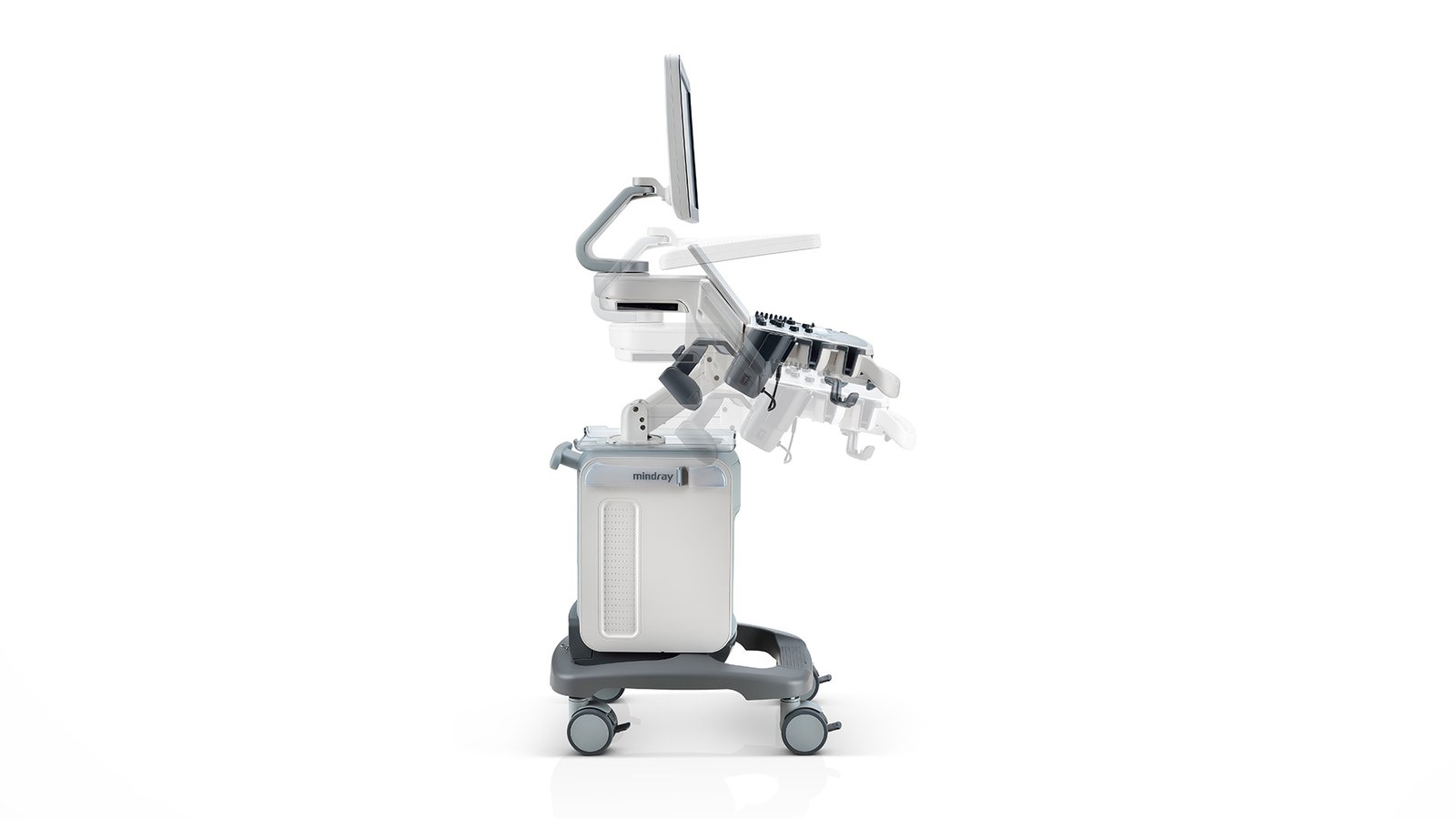
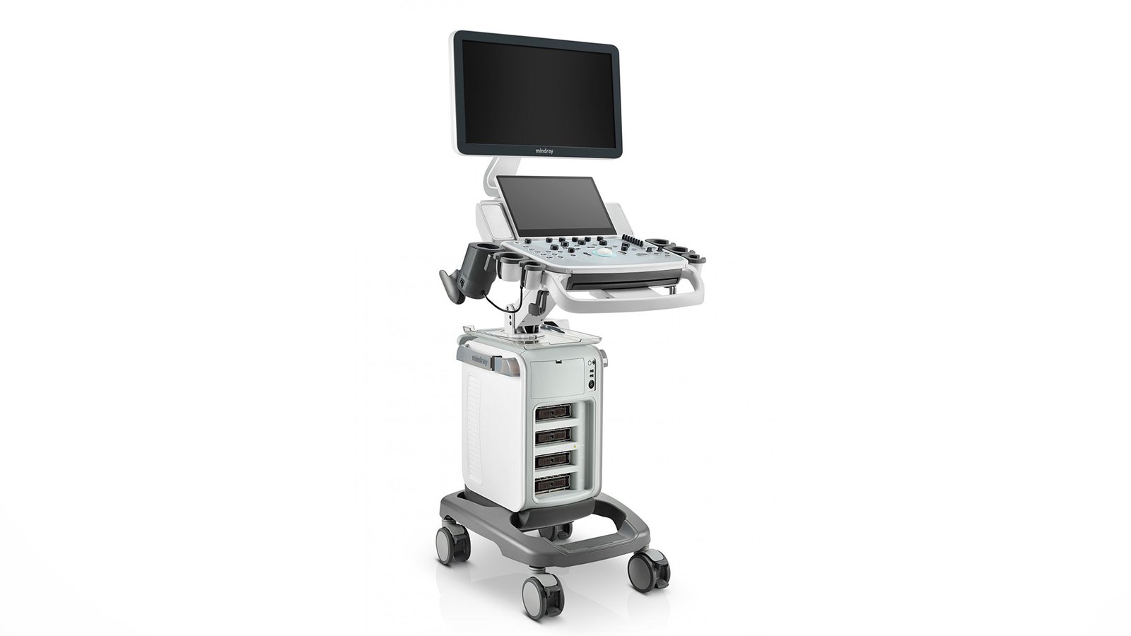
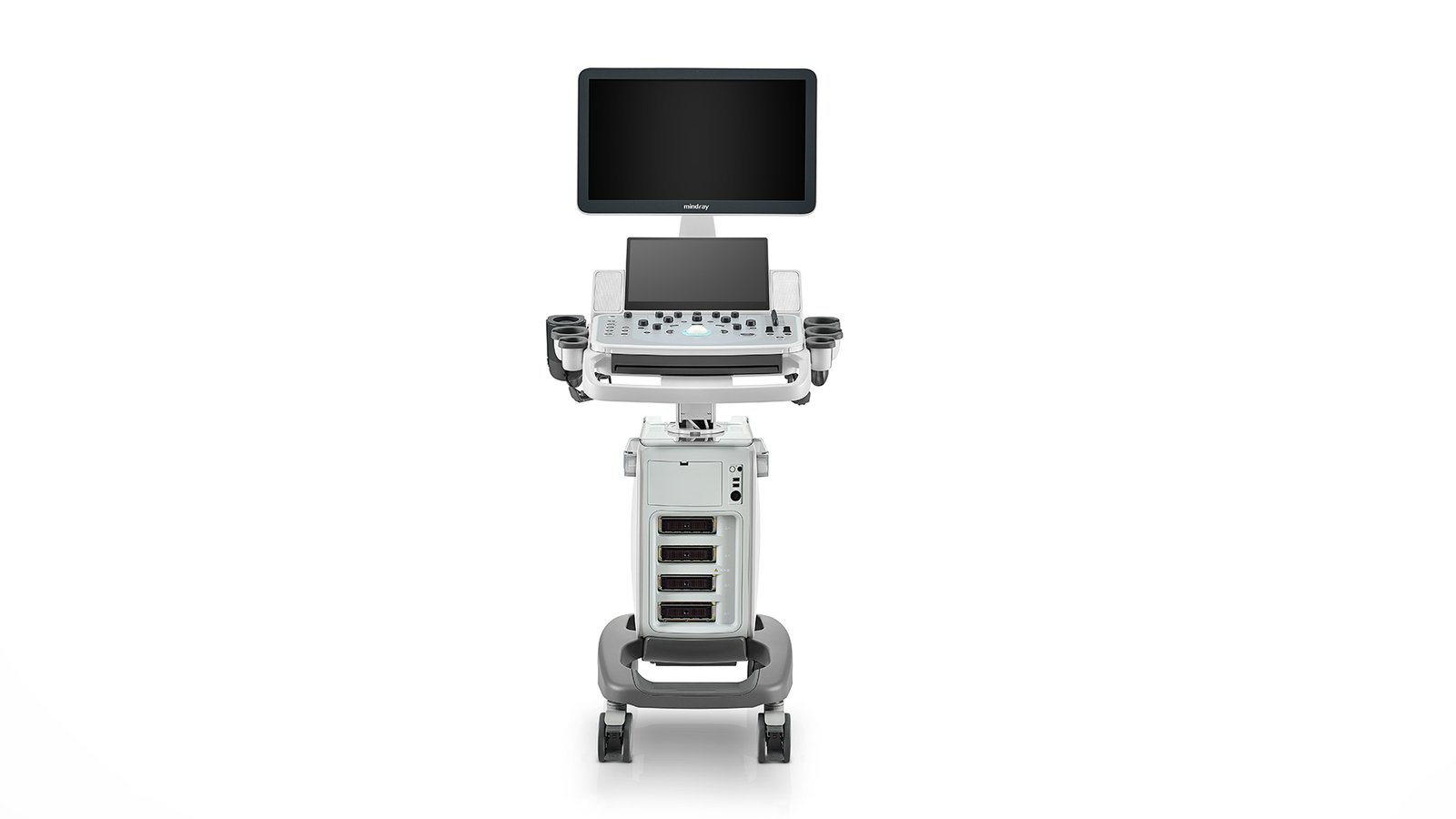
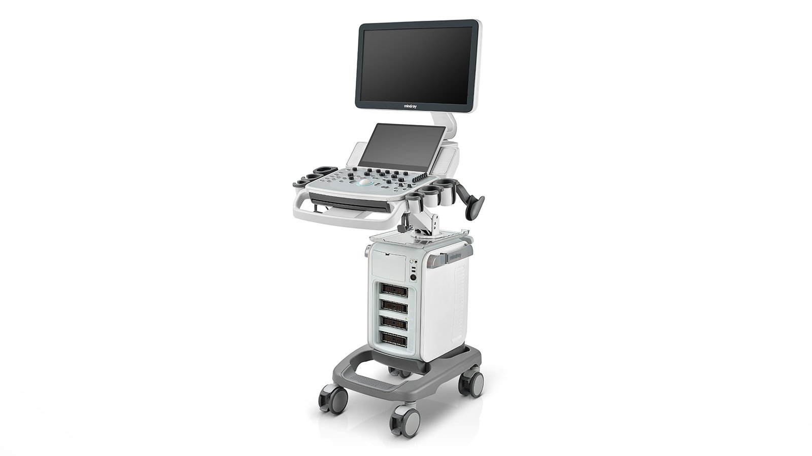


УЗИ сканер Mindray DC-40
Ultrasound scanner Mindray DC-40
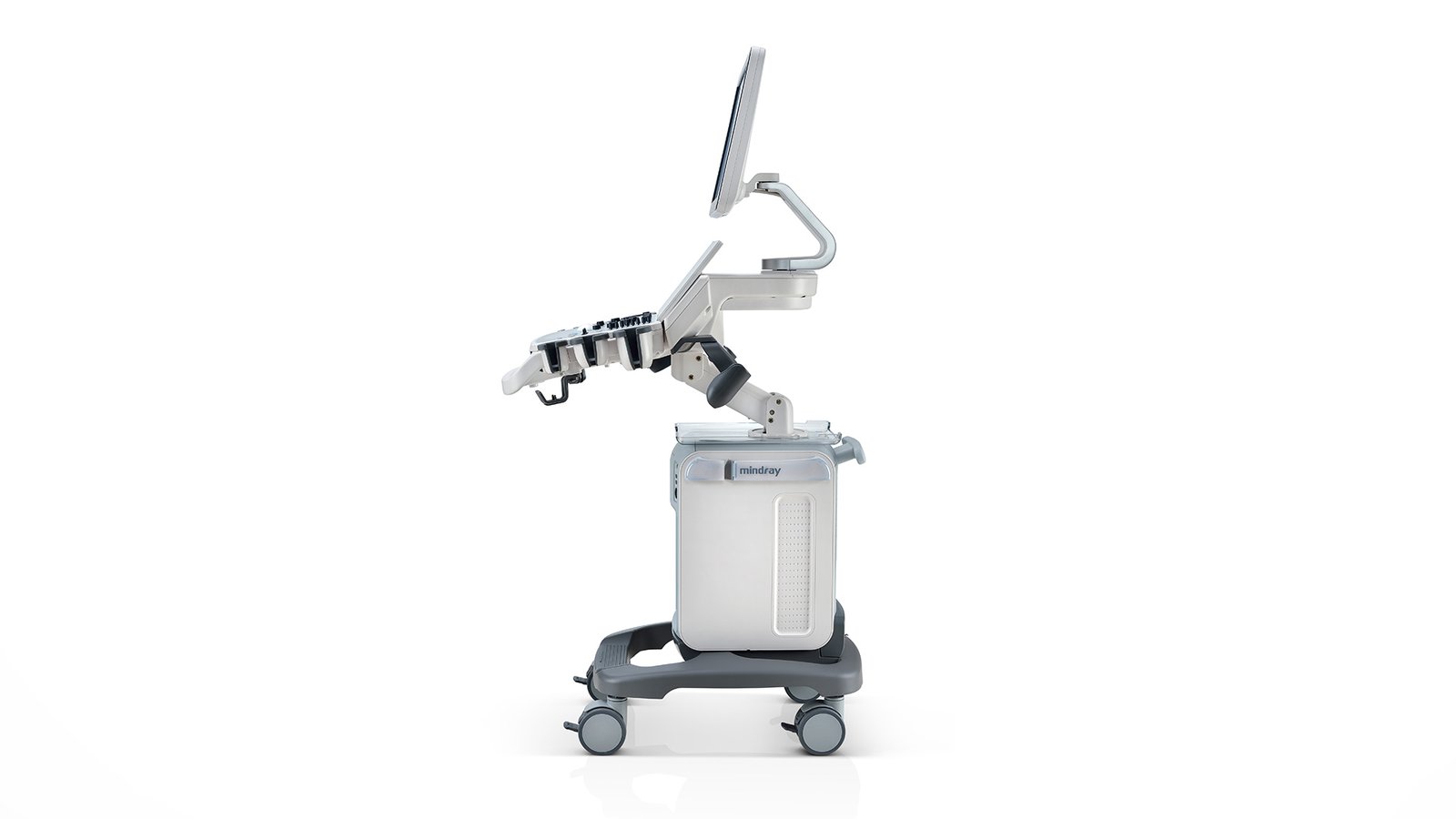
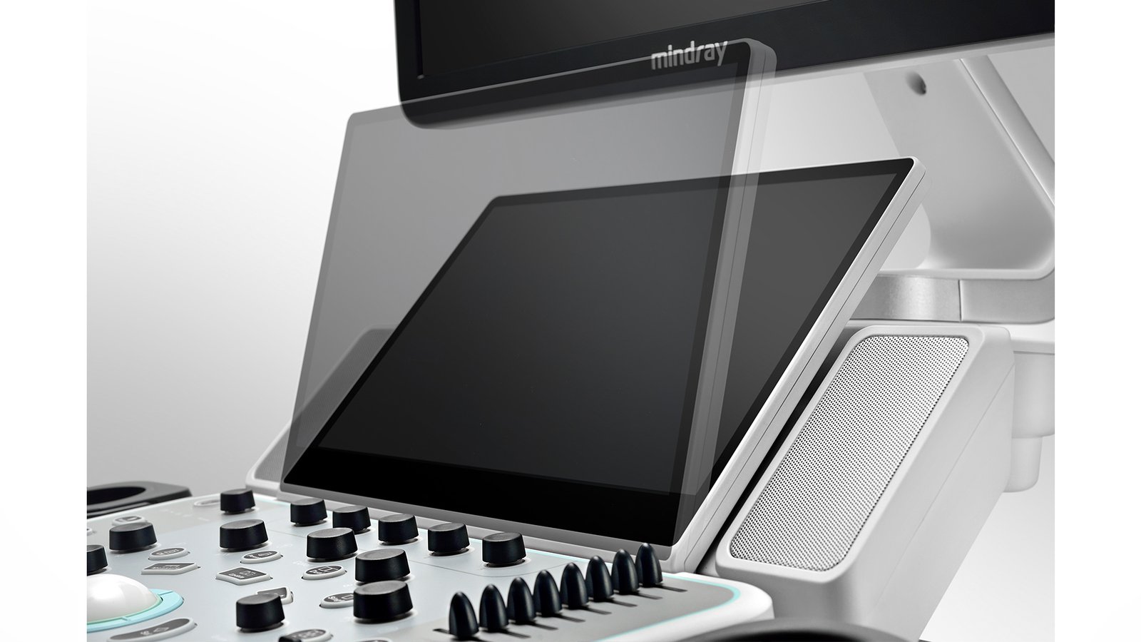
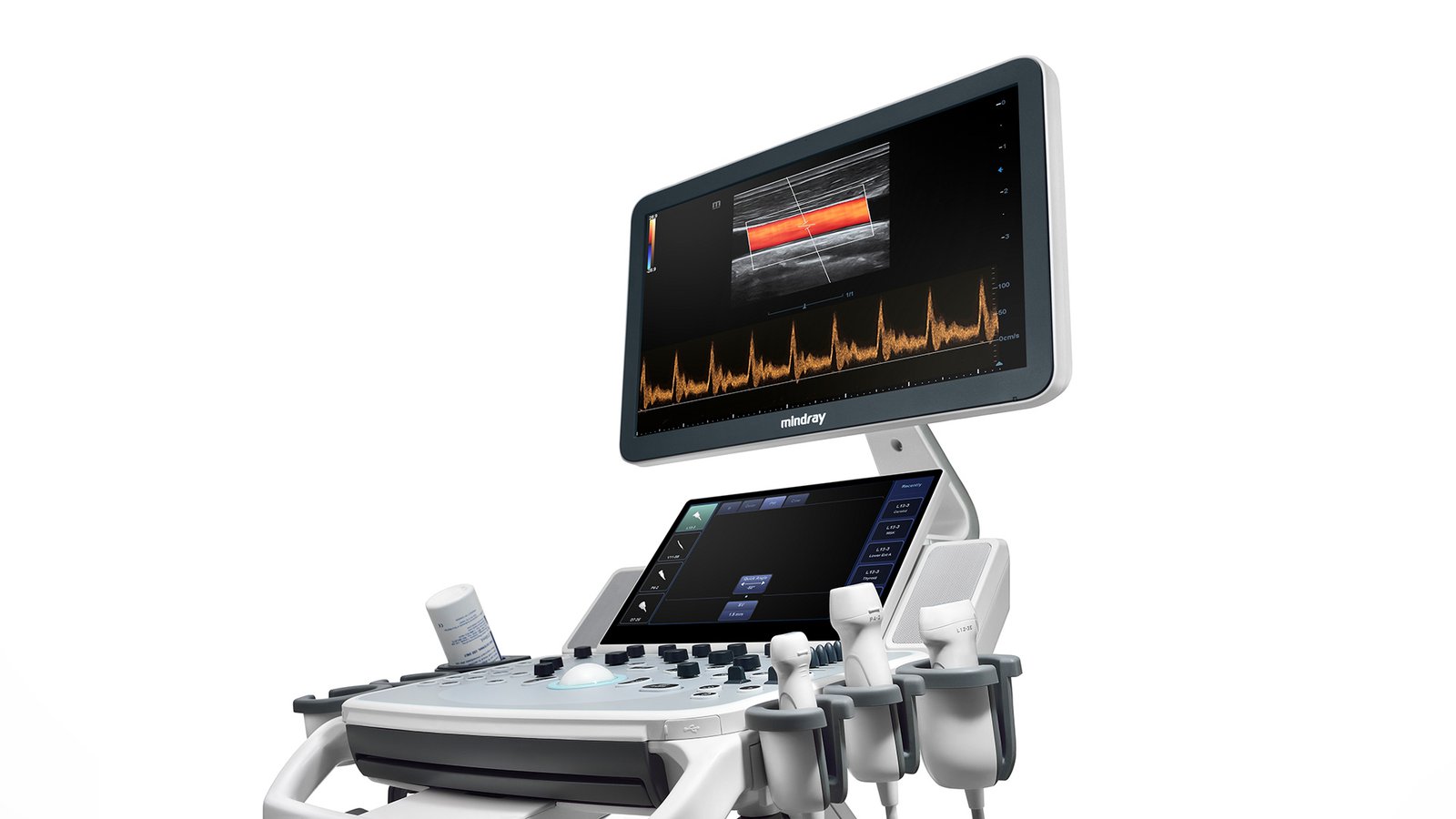
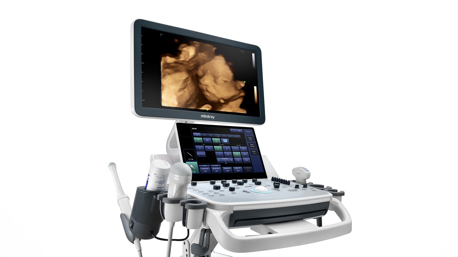
This is a new generation of equipment for ultrasound diagnostics. Mindray implements its technologies to improve the performance of healthcare workers. Portable equipment functions will resemble a phone in operation with convenient applications, technologically advanced diagnostic capabilities and high-resolution images. For greater convenience, the control panel rotates, lowers / rises in the desired direction for easy visibility. Ultrasound scanner Mindray DC-40 uses training programs, additional packages and parameters for accurate examinations, a large touch screen will reduce the burden on the doctor during increased workload, and an internal battery makes the product mobile.
Functions
- PSH - Reduces image noise for better contrast resolution and provides clarity for accurate ultrasound diagnostics.
- iBeam - different angles and fragments can be made into a single image;
- Free Xros M is the use of multiple freely placed lines for accurate, anatomically correct measurement in M mode;
- Free Xros CM - complete information about the contraction of the heart in systole and diastole with an assessment of contractility;
- iClear - Automated tissue type detection for improved quality: Uniform tissue display with clear contours;
- Multi-beam makes high-end image resolution;
- Mindray has patented Natural Touch technology, or "natural touch" in elastography, due to which seals are more effectively detected and stable reproducible results are maintained;
- Panoramic imaging with display of the required speed of the sensor and the ability to scan in two directions with iScape;
- ExFow will expand the range of diagnostics, which will allow you to see the anatomical structures in detail on all linear and convex probes;
- B-Steer improves the display of the needle, capillaries, arterioles, venules, nerve trunks and roots;
- TDI stands for Tissue Doppler Imaging.
Company developments
- iScanHelper - built-in training materials, illustrations with anatomical diagrams and echo images. Presence of normal echograms for comparison with real-time images. Scheme with the correct image of the position of the patient and the location of the sensor during the diagnosis;
- Auto IMT - the state of the carotid artery is checked by automatic measurement of wall thickness;
- Auto LV – left ventricular (LV) function is measured automatically. This is a vital function that evaluates cardiac output before pathology or with existing pathologies of the heart;
- Uncompressed data formats - the ability to process saved photos after scanning, with the addition of comments and measurement data, to improve performance during ultrasound diagnostics;
- IZoom is just one button for full screen mode;
- iTouch - image optimization with one click;
- iStorage, iMeasurement, iReport - transfer images to a computer. Software that works offline;
- iStation - operational management of patient information with integration, viewing, archiving and retrieval of information;
- MedSight - Clinical images and videos are transmitted in an interactive application from OS to IOS or Android. All research is at your fingertips.
Sensors
- convex sensor;
- Microconvex high-frequency sensor (pediatrics and neonatology);
- Linear sensor;
- Sector phased;
- intracavitary;
- Endorectal biplane.
- DICOM - Yes
- WiFi - Yes
- Auto measurements in 3D mode - No
- Automatic Image Optimization - Yes
- Automatic calculation of hemodynamic parameters from the Doppler spectrum (tracing / spectrum contouring) - Yes
- Automatic intima-media thickness (IMT) calculation - Yes
- Automatic determination of the Simpson ejection fraction - No
- Anatomical M-Mode - Yes
- Vector Blood Flow Mapping - No
- Type of elastography - Compression
- Types of supported sensors - Convex, Micro-convex, Pencil, Linear (up to 15 MHz), Linear low-frequency, Cavity convex, Cavity biplane (convex + convex), Sector phased adult, Volumetric convex, Sector phased pediatric, Linear high-frequency, Volumetric cavity
- Virtual light source in 3D - Yes
- Built-in rechargeable battery - Yes
- High Frequency Pulse Doppler - Yes
- High Sensitivity Doppler (Microvascular Imaging) - No
- Pulsed Wave Doppler - Yes
- Device class - Medium
- Number of active connectors for sensors - 4
- Number of active connectors for sensors - 4
- Command touch display - Yes
- Multi-beam scanning/compounding - Yes
- Beam tilt in Doppler modes on linear B-steer transducers - Yes
- The presence of automatic calculation of the collar space - Yes
- The presence of compression elastography - Yes
- Availability of shear wave elastography - No
- Directed ED - Yes
- Non-Doppler imaging of blood flow - No
- Volumetric imaging of the fetal heart (STIC) - No
- Volumetric scanning (4D) - Yes
- Option to obtain a three-dimensional image in the mode of color Doppler mapping (three-dimensional reconstruction of the color flow) - No
- Assessment of myocardial deformation (speckle tracking) - No
- Option Pack 5D - No
- Panoramic Scan - Yes
- Grain Suppression - Yes
- ECG Block Support - Yes
- Support for studies with contrast agents - Yes
- Support for Fusion technology (combining images on CT / MRI with ultrasound) - No
- Gel warmer - Yes
- Constant Wave Doppler (CW) - Yes
- The program for automatic measurement of the main parameters of fetal biometrics in obstetrics - Yes
- Sensor splitter for portable scanners - No
- Screen Size - ″17
- Height adjustable control panel - Yes
- Specialization (ultrasound) - General research, Gynecology and obstetrics
- Stress echocardiography - No
- Solid State Drive (SSD) - No
- Device type - Stationary
- Supported Sensor Types - High Density
- Tissue Doppler (TDI) - Yes
- Trapezoidal mode (virtual convex) - Yes
- 3D free hand reconstruction - Yes
- Needle Visualization Improvement for Linear Gauges - No
- Ultrasound tomography - Yes
- Color Doppler (CD) - Yes
- Power Doppler - Yes