УЗИ сканер Mindray DC-40
Description
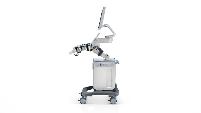
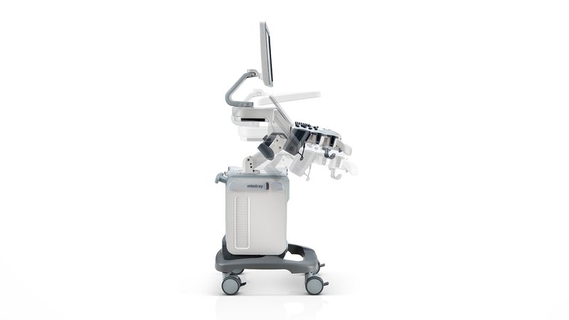
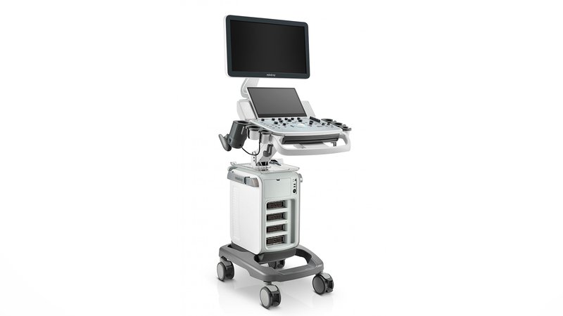
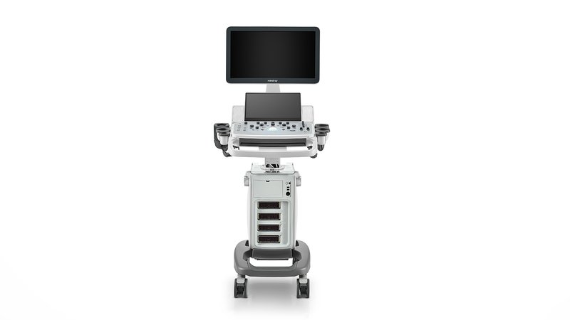
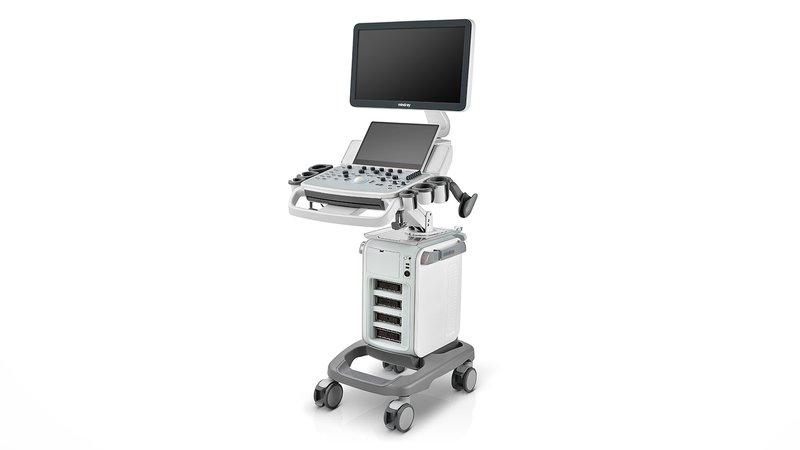
Ultrasound scanner Mindray DC-40
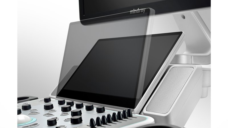
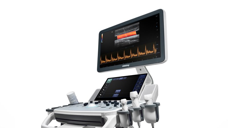
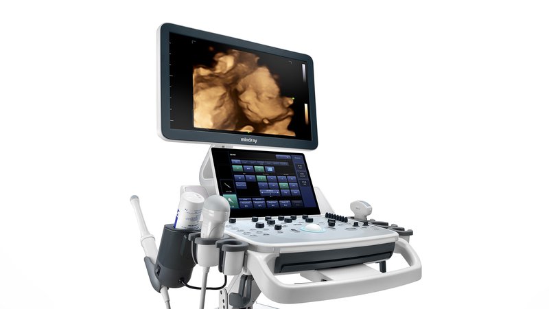
This is a new generation of equipment for ultrasound diagnostics. Mindray implements its technologies to improve the performance of healthcare workers. Portable equipment functions will resemble a phone in operation with convenient applications, technologically advanced diagnostic capabilities and high-resolution images. For greater convenience, the control panel rotates, lowers / rises in the desired direction for easy visibility. Ultrasound scanner Mindray DC-40 uses training programs, additional packages and parameters for accurate examinations, a large touch screen will reduce the burden on the doctor during increased workload, and an internal battery makes the product mobile.
Functions
- PSH - Reduces image noise for better contrast resolution and provides clarity for accurate ultrasound diagnostics.
- iBeam - different angles and fragments can be made into a single image;
- Free Xros M is the use of multiple freely placed lines for accurate, anatomically correct measurement in M mode;
- Free Xros CM - complete information about the contraction of the heart in systole and diastole with an assessment of contractility;
- iClear - Automated tissue type detection for improved quality: Uniform tissue display with clear contours;
- Multi-beam makes high-end image resolution;
- Mindray has patented Natural Touch technology, or "natural touch" in elastography, due to which seals are more effectively detected and stable reproducible results are maintained;
- Panoramic imaging with display of the required speed of the sensor and the ability to scan in two directions with iScape;
- ExFow will expand the range of diagnostics, which will allow you to see the anatomical structures in detail on all linear and convex probes;
- B-Steer improves the display of the needle, capillaries, arterioles, venules, nerve trunks and roots;
- TDI stands for Tissue Doppler Imaging.
Company developments
- iScanHelper - built-in training materials, illustrations with anatomical diagrams and echo images. Presence of normal echograms for comparison with real-time images. Scheme with the correct image of the position of the patient and the location of the sensor during the diagnosis;
- Auto IMT - the state of the carotid artery is checked by automatic measurement of wall thickness;
- Auto LV – left ventricular (LV) function is measured automatically. This is a vital function that evaluates cardiac output before pathology or with existing pathologies of the heart;
- Uncompressed data formats - the ability to process saved photos after scanning, with the addition of comments and measurement data, to improve performance during ultrasound diagnostics;
- IZoom is just one button for full screen mode;
- iTouch - image optimization with one click;
- iStorage, iMeasurement, iReport - transfer images to a computer. Software that works offline;
- iStation - operational management of patient information with integration, viewing, archiving and retrieval of information;
- MedSight - Clinical images and videos are transmitted in an interactive application from OS to IOS or Android. All research is at your fingertips.
Sensors
- convex sensor;
- Microconvex high-frequency sensor (pediatrics and neonatology);
- Linear sensor;
- Sector phased;
- intracavitary;
- Endorectal biplane.