

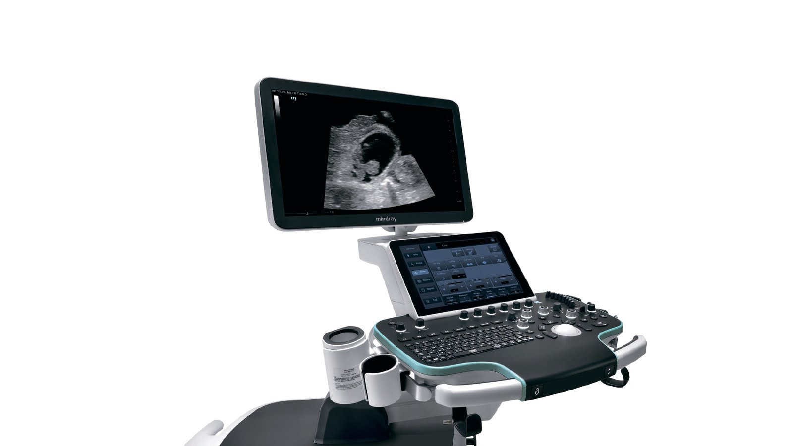
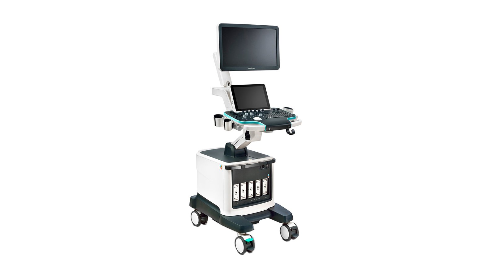


Узи аппарат Mindray Resona 6 Exp
- Manufacturer
- Mindray
Ultrasound machine Mindray Resona 6 Exp
Mindray Resona 6 Exp is in demand among professionals due to a large number of specialized functions. Works on the ZST platform, which allows for zone scanning, as well as channel data processing. Resona 6 Exp significantly improves image quality, which helps in diagnostics. It automatically optimizes the image when rendering complex areas. For example, the thyroid gland or small vessels. Recommended for use in public institutions.
Advantages
- Technology:
An innovative data processing method is channel data representation based on special ZST+ technology. This is a new stage in the development of ultrasound research technologies. There is a transformation of ultrasonic parameters, which are obtained with the traditional formation of an ultrasonic beam. ZST+ technology enables innovative solutions for high quality imaging. For example, it provides enhanced acoustic imaging as well as sound velocity compensation, dynamic pixel focusing, and improved channel data representation. Based on the fully accessible sample, diagnostic imaging is performed.
- Enhanced Acoustic Imaging
It became possible to extract more information for each frame, and the speed of imaging increased ten times than with traditional ultrasonic beam formation. This was achieved by transmitting as well as receiving signals regarding the number of large areas.
- Sound speed compensation
The device's SI system is able to intelligently select the optimal sound speed, which provides a retrospective analysis of the received channel data. Improved image fidelity for each type of fabric. Resona 6 Exp provides adjustable tissue-specific optimization.
- Pixel dynamic focus
The device provides unsurpassed uniformity of the image on all area of scanning at pixel level. When examining a patient, you no longer have to separately adjust the focal length in order to obtain a homogeneous image.
- Improved processing of channel data
This significantly improved the clarity of the visualization. Multiple retrospective processing of channel data allows effective use of acoustic information for a better image. Coherent spatial synthesis: spatial combining further improves the quality of the resulting image.
HD area: Get a clearer image within the area of interest.
ZST+ technology collects and stores raw acoustic data. Therefore, the sample-based diagnostic imaging method will allow the system to perform retrospective processing of the data, as well as change the imaging parameters of the saved images. This is essential to improve clinical performance.
Additional functions:
- RIMT
With the help of radio data, the thickness of the intima-media complex is measured. This technology is able to provide CMM thickness measurement in automatic mode. In addition, all measurements take place in real time. At the same time, they have exceptional accuracy down to 5 microns. Diagnostic accuracy also improves quantitative analysis within six cardiac cycles. In addition, there is now less dependence on image quality.
- UWN contrast imaging
Ultra-wide non-linear imaging for contrast echography allows the system to detect and exploit non-linear primary signals as well as second harmonics. Create an image of higher quality and with a greater degree of sensitivity to secondary signals. Now lower span requirements are needed.
- Smart Planes
Midray introduced a unique, groundbreaking technology that makes the Resona 6 Exp an innovative system in the world of ultrasound. It is able to provide fully automatic as well as accurate acquisition of important projections. It performs frequently used measurements of the fetal CNS. This results in improved performance as well as smart diagnostics and reduced user dependency. This technology is the most convenient tool for a specialist, as it can significantly increase the efficiency of scanning. This was achieved by increasing accuracy in combination with the full automation of labor-intensive work. Simply press a button to have the 3D data generated by the fetal brain scan provide information on standard CNS scanning planes: MSP, TCP, TTP and TVP. As these, measurements such as BDP, OG, LZR, Cisternae Major, Cerebellar Transverse Diameter and Lateral Ventricular Width are obtained.
- Natural Touch elastography method
This method is able to provide excellent sensitivity and incredible reproducibility of results regardless of experience.
- DICOM Yes
- WiFi - Yes
- Auto measurements in 3D mode - Yes
- Automatic Image Optimization - Yes
- Automatic calculation of hemodynamic parameters from the Doppler spectrum (tracing / spectrum contouring) - Yes
- Automatic intima-media thickness (IMT) calculation - Yes
- Automatic determination of the Simpson ejection fraction - Yes
- Anatomical M-Mode - Yes
- Vector Blood Flow Mapping - No
- Type of elastography - Compression, Shear wave
- Types of supported sensors - Convex, Micro-convex, Pencil, Linear (up to 15 MHz), Linear low-frequency, Cavitary convex, Sector phased adult, Volumetric convex, Intraoperative linear, Sector phased pediatric, Sector phased neonatal, Transesophageal adult, Linear high-frequency, Volumetric cavity, Linear UHF
- Virtual light source in 3D - Yes
- Built-in rechargeable battery - No
- High Frequency Pulse Doppler - Yes
- High Sensitivity Doppler (Microvascular Imaging) Yes
- Pulsed Wave Doppler Yes
- Device class - Premium
- Number of active connectors for sensors - 4
- Number of parking connectors for sensors - 1
- Command touch display Yes
- Multi-beam scanning/compounding - Yes
- Beam tilt in Doppler modes on linear B-steer transducers - Yes
- The presence of automatic calculation of the collar space - Yes
- The presence of compression elastography - Yes
- Availability of shear wave elastography - Yes
- Directed ED - Yes
- Non-Doppler imaging of blood flow - No
- Volumetric imaging of the fetal heart (STIC) - Yes
- Volumetric scanning (4D) - Yes
- Option to obtain a three-dimensional image in the mode of color Doppler mapping (three-dimensional reconstruction of the color flow) - Yes
- Assessment of myocardial deformation (speckle tracking) - Yes
- Option Pack 5D - No
- Panoramic Scan Yes
- Grain Suppression Yes
- ECG Block Support - Yes
- Support for studies with contrast agents - Yes
- Support for Fusion technology (combining images on CT / MRI with ultrasound) - No
- Gel warmer - Yes
- Constant Wave Doppler (CW) Yes
- The program for automatic measurement of the main parameters of fetal biometrics in obstetrics - Yes
- Sensor splitter for portable scanners - No
- Screen size ″21
- Specialization (ultrasound) - General research, Cardiology
- Stress echocardiography - Yes
- Solid State Drive (SSD) Yes
- Device type - Stationary
- Supported Sensor Types - High Density, Monocrystalline, Matrix
- Tissue Doppler (TDI) Yes
- Trapezoidal mode (virtual convex) - Yes
- 3D free hand reconstruction Yes
- Needle Visualization Improvement for Linear Gauges - Yes
- Color Doppler (CD) Yes
- Power Doppler Yes