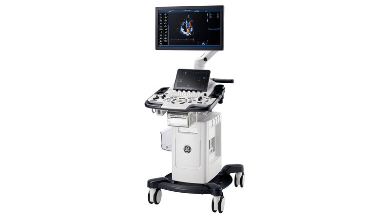
УЗИ сканер GE Healthcare Vivid T9
- Manufacturer
- GE
GE Vivid T9
Universal ultrasound machine for cardiology and general examinations in adults and children, with advanced diagnostic capabilities in obstetrics and gynecology, angiology, urology, examination of the abdominal organs, small organs, surface structures and the musculoskeletal system, urology, transcranial and transesophageal studies. The console is equipped with a 21.5-inch LCD monitor. The versatile Vivid T9 system delivers superior image quality across a wide range of exams and can be customized to meet institutional needs. Ease of use allows you to effectively use the Vivid T9 R1 cardiovascular ultrasound console in any environment: due to the simple interface of the Vivid line and the presence of a touch panel. Additional options (StressEchoCG package, AutoEF 2.0, AFI 2.0 and an automated research protocol) expand the range and efficiency of using the console. GE's patented "raw data" technology enables additional post-processing of stored data and advanced quantitative analysis. Smart Standby mode automatically saves all study data in the event of an emergency shutdown or power failure. After power is restored, the system returns to the state in which the automatic shutdown occurred.
Advantages
- Simple and reliable scanning
The system is designed to provide ease of operation and transportation in various conditions. Its intuitive user interface is the console itself, with all the benefits of its applications, features, streamlined workflow, reliability and ease of use;
- Convenient layout of controls
The location of the touch screen, rotary controls and function buttons is convenient for the user, and all mode buttons are collected in one place next to the trackball;
- Scan coach
An overview of the main scanning methods with the image of the sensor, a schematic representation of the anatomical structures and examples of clinical images;
- Adjustable console
Adjust the console for the optimal scanning position;
- Exceptional mobility
With a light weight of only 58 kg, as well as strong wheels and front and rear handles, the system is easy to move over tiles and carpets;
- Intelligent standby
In the event of an accidental shutdown or power failure, or simply transporting the scanner to another location, the system automatically saves data and enters "Standby" mode. After power is restored, the system immediately automatically turns on, returning to the same state it was in before it was turned off;
- Wide range of sensor connections
The system has four ports for RS-type probes and four standard probe holders, as well as two additional probe ports;
- Data transfer
DICOM standards for pediatrics. Support for SR Standards Pediatric measurements sent by SR automatically populate a pediatric report at the receiving end for fast and accurate review anywhere. Improved support for DICOM SR standards for the cardiovascular system, including user-defined measurements. Advanced DICOM Viewing - Reduce time spent viewing and reporting images by using contrast, brightness, and zoom/pan controls to optimize DICOM images. Raw data transfer - transfer of user-selectable raw data in a DICOM environment;
- Safety
The design and configuration of the Vivid T9 ultrasound system is reliable and safe. LDAP - Protect patient data with LDAP (Lightweight Directory Access Protocol): Your institution's IT department will have greater control over user access to the system, helping to reduce the risk of intrusion. Customizable Login Passwords - Login and internal passwords can be customized in any way to meet the security requirements of your institution's IT department. Drive Encryption Encrypting the drive that stores archived patient data and related images keeps them secure and confidential even in the event of theft. Windows 10 operating system with lists of allowed applications to prevent unauthorized programs from running and potentially harming the scanner;
Sensors
GE's advanced sensor technology helps deliver exceptional image quality. It is used for a wide range of sensors designed to meet the needs of various studies; When designing the Vivid T9 system, our main focus was to ensure reliability. During the creation and detailed testing of the system, we focused on ensuring reliable operation even in harsh and difficult conditions. It is exceptionally resilient and can handle stress even during intense ultrasonic practice; Even after many standard on/off cycles, repeated forced shutdown episodes, and multiple probe connections/disconnections, the system error rate remained low.
Scan modes
- B-mode;
- M-mode;
- CDC;
- ED;
- 2D;
- Pulsed Wave Doppler (PW);
- TVI - tissue doppler;
- Color M-mode;
- Anatomical M-mode;
- Anatomical M-mode along a curved line (optional);
- B-Flow™/B-Flow color - visualization of hemodynamics (option).
DICOM - Yes
WiFi - Yes
Auto measurements in 3D mode - No
Automatic Image Optimization - Yes
Automatic calculation of hemodynamic parameters from the Doppler spectrum (tracing / spectrum contouring) - Yes
Automatic intima-media thickness (IMT) calculation - Yes
Automatic determination of the Simpson ejection fraction - Yes
Anatomical M-Mode - Yes
Vector Blood Flow Mapping - No
Veterinary - No
Type of elastography - No
Types of supported sensors - Linear, Convex, Microconvex, Pencil, Linear low-frequency, Cavitary convex, Sector phased adult, Intraoperative linear, Sector phased pediatric, Sector phased neonatal, TEE adult, TEE pediatric
Virtual light source in 3D - No
Battery life of portable scanners (hour) - No
Built-in rechargeable battery - No
High Frequency Pulse Doppler - Yes
High Sensitivity Doppler (Microvascular Imaging) - No
Pulsed Wave Doppler - Yes
Device class - High
Number of active connectors for sensors - 4
Number of parking connectors for sensors - No
Command touch display - Yes
Multi-beam scanning/compounding - Yes
Beam tilt in Doppler modes on linear B-steer transducers - Yes
The presence of automatic calculation of the collar space - No
Directed ED - Yes
Non-Doppler imaging of blood flow - Yes
Volumetric imaging of the fetal heart (STIC) - No
Volumetric scanning (4D) - No
Option to obtain a three-dimensional image in the mode of color Doppler mapping (three-dimensional reconstruction of the color flow) - No
Assessment of myocardial deformation (speckle tracking) - Yes
Option Pack 5D - No
Panoramic Scan - Yes
Grain Suppression - Yes
ECG Block Support - Yes
Support for studies with contrast agents - Yes
Support for Fusion technology (combining images on CT / MRI with ultrasound) - No
Gel warmer - No
Constant Wave Doppler (CW) - Yes
The program for automatic measurement of the main parameters of fetal biometrics in obstetrics - No
Sensor splitter for portable scanners - No
Screen Size - ″21
Height adjustable control panel - Yes
Specialization (ultrasound) - Cardiology
Stress echocardiography - Yes
Solid State Drive (SSD) - No
Device type - Stationary
Supported Sensor Types - High Density
Tissue Doppler (TDI) - Yes
Trapezoidal mode (virtual convex) - Yes
3D reconstruction by "free hand" method - No
Needle Visualization Improvement for Linear Gauges - Yes
Color Doppler (CD) - Yes
Power Doppler - Yes


