

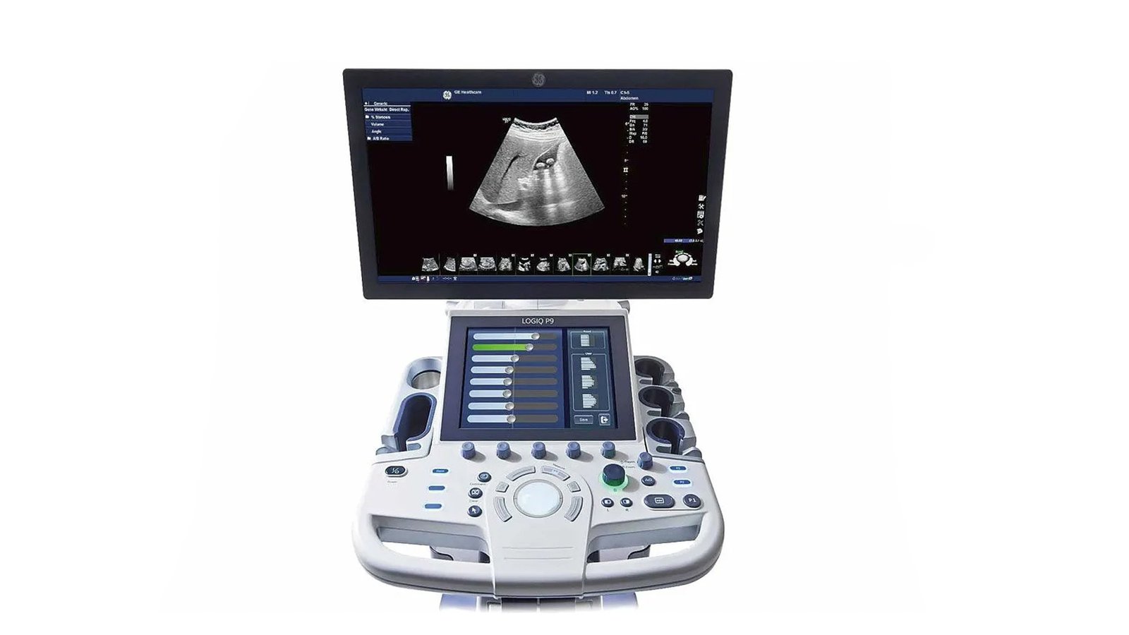


Узи сканер GE Healthcare Logiq P9
- Manufacturer
- GE
GE Logiq P9
An advanced universal type ultrasound machine manufactured by General Electric, which allows you to achieve a high-quality image. It is distinguished by high-density sensors and the most advanced hardware and software resources, giving the doctor the opportunity to carry out diagnostics in the required area of research promptly and at a professional level.
Advantages
- B-Mode, M-Mode, PW Doppler, Color Doppler and Power Doppler, Coded Radiation, Coded Tissue Harmonic;
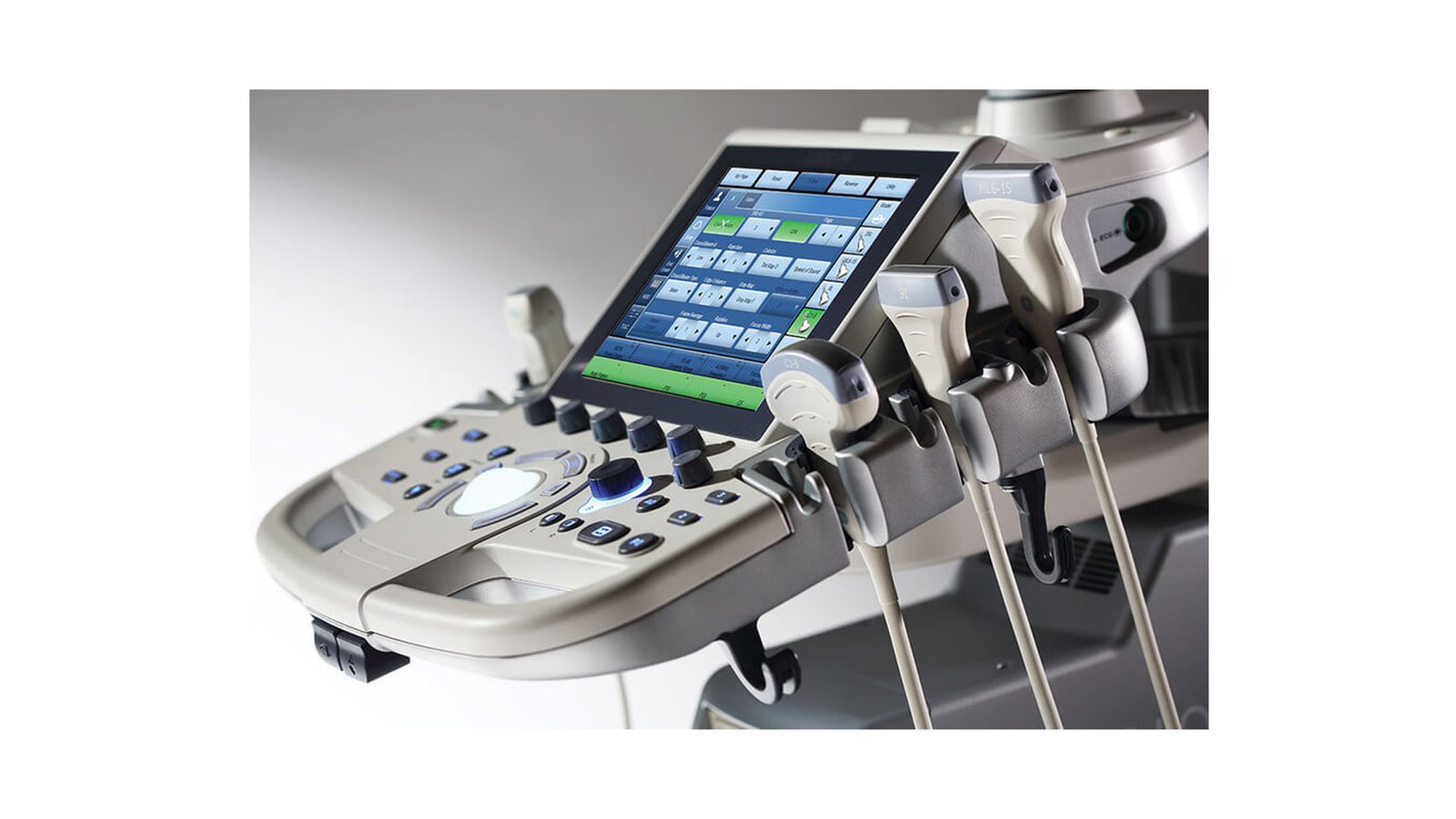
- 21.5" high resolution LCD monitor;
- Ergonomic user interface with 10.4" color touch panel;
- 4 functioning sensor connectors (high cabinet) plus 1 CW sensor connector;
- Built-in hard drive 500 GB;
- CD-R/DVD-R drive for burning CDs;
- Automatic image optimization in B-mode (ATO), spectral Doppler mode;
- CrossXBeam - scanning mode using compounding technology;
- SRI - organ-specific high-resolution imaging mode;
- 3D reconstruction program;
- Virtual convex scanning, expanding the field of view;
- Management of the built-in archive of images and data of patients;
- Measurement and reporting programs for all areas of application;
- Automatic Doppler calculations in real time;
- Virtual trainer;
- Power cable.
Additional software and hardware options
- B-Flow technology for high-precision hemodynamic imaging;
- Compare Assistant for comparing and comparing current and previously acquired images;
- Measure Assistant Breast - automatic contouring and measurement of formations in the mammary gland;
- Measure Assistant OB - automatic fetal biometry;
- LOGIQ View - panoramic scanning;
- Coded Contrast Imaging for examination with contrast agents;
- DICOM 3 - data transfer protocol;
- Scan Assistant - automated research protocols;
- Real Time 4D - real-time volumetric scanning mode (includes inversion mode, cine loop, 3D in color flow mode);
- VOCAL II - calculation program when using 4D;
- VCI - volumetric contrast image mode;
- TUI - Tomographic ultrasound;
- Elastography - elastography;
- Quantification Elastography - Quantitative elastography;
- CW - constant wave doppler;
- TVI - tissue doppler;
- Auto-IMT - automatic calculation of intima-media;
- Auto EF - program for automatic assessment of the global contractile function of the left ventricle;
- Gel warmer.
- Screen Size - ″21
- DICOM Yes
- Number of active connectors for sensors - 4
- Command touch display Yes
- Volumetric scanning (4D) - Yes
- 3D free hand reconstruction Yes
- Automatic calculation of intima-media thickness (IMT) - Yes
- Trapezoidal mode (virtual convex) - Yes
- Panoramic Scan Yes
- Needle Visualization Improvement for Linear Gauges - Yes
- Ultrasound tomography - Yes
- The program for automatic measurement of the main parameters of fetal biometrics in obstetrics - Yes
- The presence of automatic calculation of the collar space - No
- Option Pack 5D - No
- Constant Wave Doppler (CW) Yes
- Color Doppler (CD) Yes
- Tissue Doppler (TDI) Yes
- Anatomical M-Mode - Yes
- Support for Fusion technology (combining images on CT / MRI with ultrasound) - No
- Volumetric imaging of the fetal heart (STIC) - Yes
- ECG Block Support - Yes
- Support for studies with contrast agents - Yes
- Device type - Stationary
- Special Sensor Support - Matrix
- Specialization (ultrasound) - General studies
- Device class - High
- Veterinary - No
- Type of elastography - Compression
- Built-in rechargeable battery - No
- Gel warmer - No
- Number of parking connectors for sensors - No
- Sensor splitter for portable scanners - No
- Battery life of portable scanners (hour) - No
- Assessment of myocardial deformation (speckle tracking) - Yes
- Solid State Drive (SSD) - No
- Height adjustable control panel - Yes
- Pulsed Wave Doppler Yes
- High Frequency Pulse Doppler - Yes
- Power Doppler Yes
- Directed ED - Yes
- Vector Blood Flow Mapping - No
- Automatic determination of the Simpson ejection fraction - Yes
- Automatic Image Optimization - Yes
- Automatic calculation of hemodynamic parameters from the Doppler spectrum (tracing / spectrum contouring) - Yes
- Non-Doppler imaging of blood flow - Yes
- Stress echocardiography - Yes
- WiFi - Yes
- Beam tilt in Doppler modes on linear B-steer transducers - Yes
- Multi-beam scanning/compounding - Yes
- Grain Suppression Yes
- Virtual light source in 3D - No
- Option to obtain a three-dimensional image in the mode of color Doppler mapping (three-dimensional reconstruction of the color flow) - No
- Auto measurements in 3D mode - Yes
- High Sensitivity Doppler (Microvascular Imaging) - No
- Supported Sensor Types - High Density, Matrix
Types of supported sensors
Linear, Convex, Microconvex, Pencil, Linear low-frequency, Cavitary convex, Cavitary biplane (convex + convex), Sector phased adult, Volumetric convex, Intraoperative linear, Sector phased pediatric, Sector phased neonatal, Linear high-frequency, Volumetric cavitary, Linear ultra-high frequency