


УЗИ система GE Healthcare Venue 50
- Manufacturer
- GE
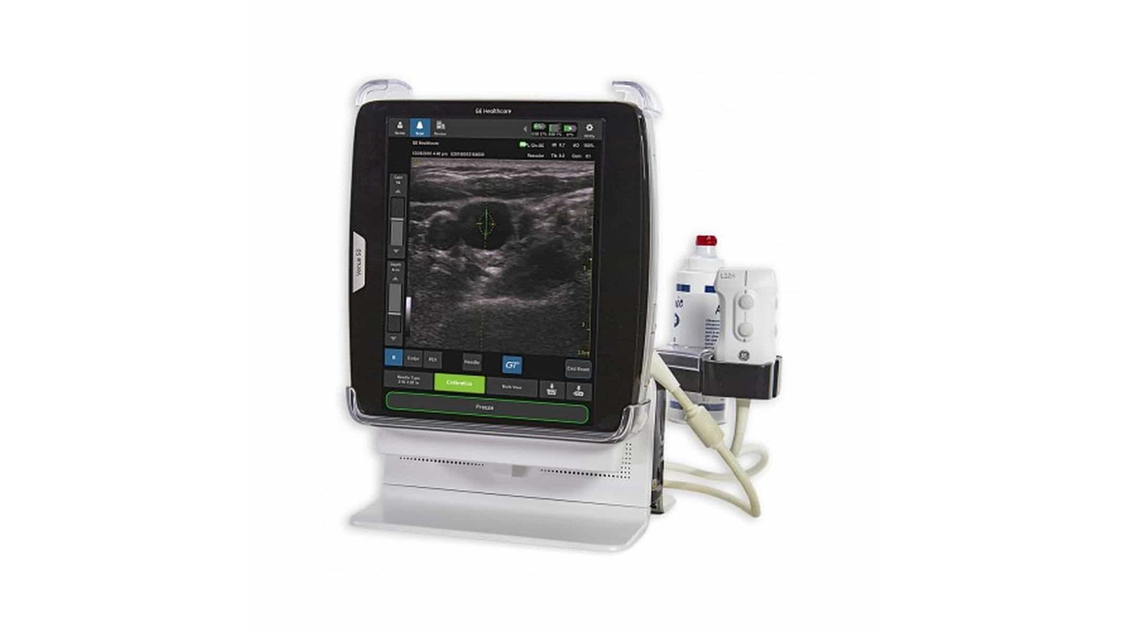
Ultrasound system GE Healthcare Venue 50
High performance flatbed ultrasound scanner with easy to use touch screen and improved needle visualization. The system is characterized by high loading speed and readiness for operation in 16 seconds, the presence of advanced imaging tools, ease of operation and clinical accuracy of the results obtained. The Venue 50 is designed for ultrasound imaging, measurements and analysis of anatomical organs in a range of clinical applications, including: The sleek and portable design of the Venue 50 fits perfectly into tight spaces with virtually no space required. The seamless design allows easy cleaning and disinfection of the system. Flexible data management and optional DICOM connectivity help expedite image storage and archiving at the point of care or at the patient's bedside.
Advantages
- Results in one touch. Possibility to select sensor and preset parameters in one step;
- Tablet-like navigation. Ability to use on-screen gestures during the procedure: squeeze, slide and touch - even when wearing gloves;
- Quick turn on. You won't even have time to put on your gloves before the system is ready to go;
- Mobility and wireless connectivity. The battery-operated system can be moved from one patient to another without turning off the power;
- Start imaging immediately after applying the gel. Get sharp images without time-consuming detailed adjustments.
Areas of use
- Intraoperative studies;
- Examination of the chest and pleural cavity for movement and fluid recognition, visual control of interventional procedures (tissue biopsy, fluid drainage, vascular access, non-vascular procedures);
- Abdominal, gynecological and urological examinations, including intracavitary, fetal and obstetric examinations; pediatric and neonatal research; examinations of superficially located organs, transcranial examinations of newborns and adults, cardiac examinations; vascular studies; studies of the musculoskeletal system.
Standard parameters
- System boot 16 s;
- ATO - automatic image optimization;
- CrossXBeam - compounding;
- Scaling (Zoom);
- Splitting the screen into two parts (Split);
- Cine loop 250 MB;
- Saving information in JPEG and MPEG4 format to SD-card or USB-drive;
- Measurement and calculations, editing;
- Connector for connection to the docking station;
- DVI output for connecting a second monitor;
- USB cable;
- SD card and card reader;
- Battery.
Accessories
- Card reader for SD cards;
- SD card;
- USB cable;
- USB flash drive 4GB;
- USB wireless adapter;
- Battery for Venue 50;
- USB barcode reader;
- Protective cover for the operating room;
- Power module.
- DICOM - Yes
- WiFi - Yes
- Auto measurements in 3D mode - No
- Automatic Image Optimization - Yes
- Automatic calculation of hemodynamic parameters from the Doppler spectrum (tracing / spectrum contouring) - No
- Automatic calculation of the thickness of the intima-media complex (IMT) - No
- Automatic determination of the Simpson ejection fraction - No
- Anatomical M-Mode - No
- Vector Blood Flow Mapping - No
- Veterinary - No
- Type of elastography - No
- Types of supported sensors - Linear, Convex, Microconvex, Cavity Convex, Sector Phased Adult, Intraoperative Linear
- Virtual light source in 3D - No
- Battery life of portable scanners (hour) - 1
- Built-in rechargeable battery - Yes
- High Frequency Pulse Doppler - No
- High Sensitivity Doppler (Microvascular Imaging) - No
- Pulsed wave doppler - No
- Device class - High
- Number of active connectors for sensors - 1
- Number of parking connectors for sensors - No
- Command touch display Yes
- Multi-beam scanning/compounding - Yes
- Beam tilt in Doppler modes on linear B-steer transducers - Yes
- The presence of automatic calculation of the collar space - No
- Directed ED - No
- Non-Doppler imaging of blood flow - No
- Volumetric imaging of the fetal heart (STIC) - No
- Volumetric scanning (4D) - No
- Option to obtain a three-dimensional image in the mode of color Doppler mapping (three-dimensional reconstruction of the color flow) - No
- Assessment of myocardial deformation (speckle tracking) - No
- Option Pack 5D - No
- Panoramic Scan - No
- Grain Suppression - Yes
- ECG block support - No
- Support for studies with contrast agents - No
- Support for Fusion technology (combining images on CT / MRI with ultrasound) - No
- Gel warmer - No
- Constant Wave Doppler (CW) - No
- The program for automatic measurement of the main parameters of fetal biometrics in obstetrics - No
- Sensor splitter for portable scanners - Yes
- Screen Size - ″12
- Control panel height adjustment - No
- Specialization (ultrasound) - General Research
- Stress echocardiography - No
- Solid State Drive (SSD) - No
- Device type - Tablet
- Types of sensors supported - No
- Tissue Doppler (TDI) - No
- Trapezoidal mode (virtual convex) - No
- 3D reconstruction by "free hand" method - No
- Needle Visualization Improvement for Linear Gauges - Yes
- Color Doppler (CD) - Yes
- Power Doppler - Yes