


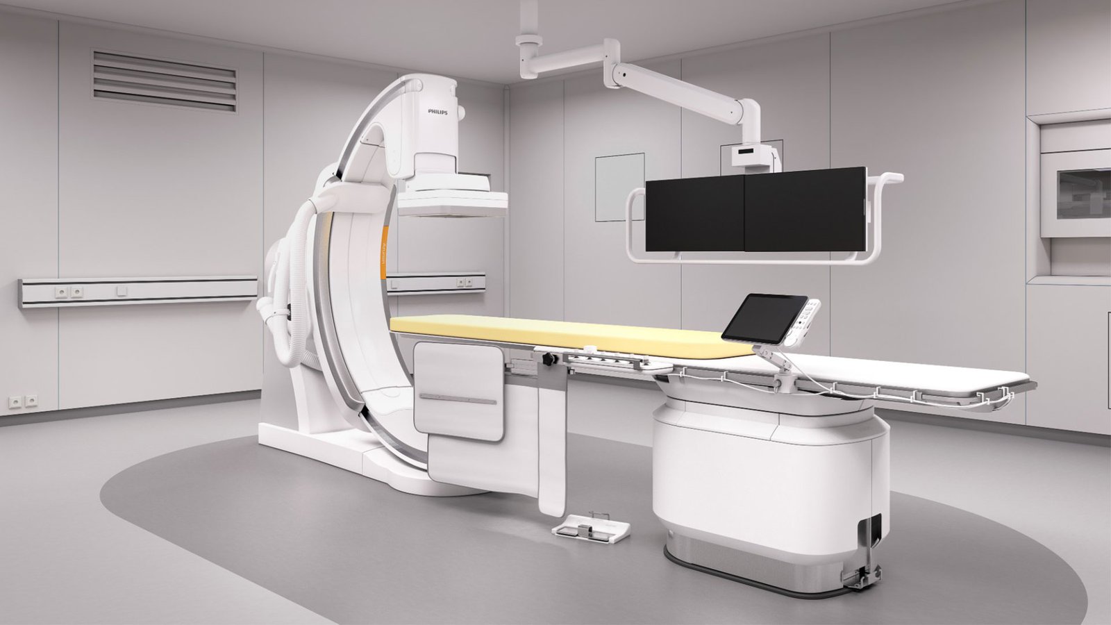


Azurion 3 M15
Angiographic system Azurion 3 M15
The Philips Azurion 3 with 15” detector is a high-tech system that enables professionals to perform complex invasive cardiac exams with superior consistency and efficiency thanks to an optimized detector size specifically designed for cardiac applications.
Advantages
- Deeper analysis
The new 15" flat detectors provide high resolution imaging and a large field of view. It is possible to visualize the aortic valve and a significant part of the aortic arch or the entire tree of coronary vessels on one screen. Thanks to the compact design, all kinds of projection angles can be achieved, including spider projection.
- Get more done
Designed specifically to save time, the Instant Parallel Working concept allows a team of specialists to perform all activities in the study room and control room without interrupting each other. For example, during a fluoroscopy or X-ray procedure, control room staff may review previous images for a patient, prepare the next examination, or complete a reporting procedure for another patient.
- Informative real-time visual control
Medical images and information highlighted on a black background of the user interface are easy to read. Users can operate the system intuitively with prominent buttons. All Azurion systems use the same standardized user interface, which reduces training time and eases the transition of medical personnel from one laboratory to another.
- Possibility of modernization
The Azurion system infrastructure is designed to be upgradeable and expandable. This standardized appliance platform provides access to a new generation of connected medical applications and technologies. As new needs arise and requirements change, you can easily integrate additional third-party features and applications.
- Floor mounting type C-shaped tripod;
- Movement of the C-shaped tripod with the help of an electric drive and manually;
- C-arm travel interval 270 deg.
- The maximum speed of rotation of the C-arm (in degrees / sec.), not less than 25;
- Three-axis positioning. Availability of additional positions of the system, programmed by the user;
- The presence of a system to protect the patient and staff from injuries when moving various components of the system;
- The presence of a mechanism to prevent the patient from colliding with the moving parts of the angiosystem, which includes an alarm in case of dangerous proximity and touch sensors that stop the movement of the system.
- Ability to move along 8 axes
- 27% reduction in patient pre-positioning time
- Size of the maximum field of view of the detector (diagonal): 30 or 38 cm
- Maximum power: 100 kW;
- Voltage range: 40 - 125 kV;
- Radiography current range: 1 – 1250 (in mA);
- Pixel matrix, not less than 1024x1024. Pixel size (in microns), no more than 154;
- Maximum image recording rate (frames/sec.), not less than 30;
- Number of flat black-and-white LCD monitors on the ceiling suspension in the operating room: 2;
- Number of flat color LCD monitors on the ceiling suspension in the operating room: 1;
- Support for DICOM 3.0 protocol;
- Possibility of bidirectional exchange of patient data between the angiographic system and the local network of medical facilities using the DICOM 3.0 protocol.
- Independent multimodal workstation with the possibility of archiving on CDs and DVDs;
- Color LCD monitor with a diagonal;
- Support for DICOM 3.0 medical data transfer protocol, including printing;
- Burn images to CD/DVD in DICOM format.