


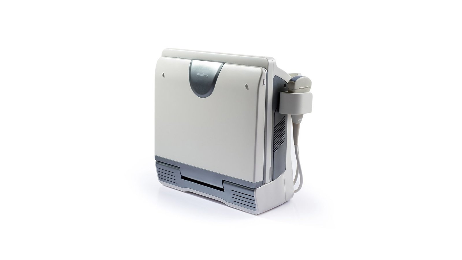
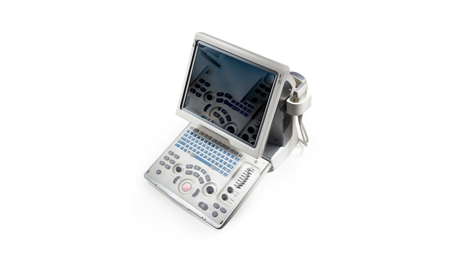


Портативный УЗИ сканер Mindray DP-50
- Manufacturer
- Mindray
Portable ultrasound scanner Mindray DP-50
Ultrasound scanner, which is a new generation of ultrasound systems from the manufacturer Mindray, which develops advanced medical equipment. Mindray DP-50 ultrasound machines are portable and developed on the unique X-treme Engine platform as a result of numerous researches. The scanner is used to examine superficial organs and structures, internal organs, limbs, and it is also possible to examine the structural features of blood vessels. The device implements advanced features and modern technologies, which makes it one of the best mobile b/w ultrasound diagnostic devices. The Mindray DP-50 ultrasound machine is equipped with high technological qualities and visualization tools, which will help medical personnel to obtain high-quality high-resolution images. The scanner handles any clinical situation with ease thanks to its streamlined design, small size and user-friendly interface for quick use. The equipment has a built-in black-and-white display using the "pseudo-color" option instead of color, which made it possible to keep the cost of the product at an affordable level for most buyers. If necessary, the device can be folded, which will make its transportation not only convenient, but also safe for the case itself.
Original Features
- Availability of a unique PSHI function, which will provide an image of high definition and contrast resolution with the possibility of noise reduction;
- iBeam technology that allows you to compose images taken from different angles to increase resolution and improve the quality of data display;
- iClear automatically recognizes structure and improves quality, resulting in sharper edges and contours, smoother and more uniform fabric details, and reduced graininess;
- An additional iScape option will give a complete overview of the anatomical structures through the panorama. Facilitates the process, makes it consistent, thanks to the combination of a speed indicator and a forward or reverse scan function;
- Multi-touch formation parameter. The signal processing speed from a single beam is increased up to four times, which makes it possible to achieve excellent image quality and a high frame rate;
- Ability to scan in various modes: B/2B/4B/M/B+M;
- The ability to tilt the display for efficient viewing;
- The presence of TGC (8 levels) - is able to optimize the image by compensating for deep tissue signals. Through the regulation of the signal of a particular area, a balanced picture can be obtained;
- ExFOV - with the help of linear and convex sensors, you can get full diagnostic information (detailed display of the anatomical structure);
- B Steer will provide the best ultrasound beam maneuver to increase the visibility of the needle, fibers and vessels. The function can be appreciated by the medical staff of your company when performing a biopsy;
- AutoIMT will allow you to find out accurate information about the carotid artery, automatically measure the thickness of the anterior and posterior walls;
- The device has 2 built-in active ports for sensors.
Developments
- The iStorage option allows you to transfer the scan and detailed information about the patient via a network cable;
- iZoom will provide instant inclusion of full-screen mode (full-screen version) with the click of a button without losing quality and level of detail;
- iStation is a technology that will allow you to quickly and efficiently manage patient information with the ability to integrate, view, archive and retrieve information;
- iTouch - allows you to optimize the image with a single click.
Sensors
- convex;
- Microconvex;
- 2 linear sensors;
- high frequency;
- In-band;
- Biplane endorectal probe.
DICOM - Yes
WiFi - No
Auto measurements in 3D mode - No
Automatic Image Optimization - Yes
Automatic calculation of hemodynamic parameters from the Doppler spectrum (tracing / spectrum contouring) - Yes
Automatic intima-media thickness (IMT) calculation - Yes
Automatic determination of the Simpson ejection fraction - No
Anatomical M-Mode - No
Vector Blood Flow Mapping - No
Types of supported sensors - Convex, Micro-convex, Linear (up to 15 MHz), Linear low-frequency, Cavity convex, Cavity biplane (convex + convex)
Virtual light source in 3D - No
Battery life of portable scanners (hour) - 2
Built-in rechargeable battery - Yes
High Frequency Pulse Doppler - No
High Sensitivity Doppler (Microvascular Imaging) - No
Pulsed Wave Doppler - Yes
Device class - Initial
Number of active connectors for sensors - 2
Number of active connectors for sensors - 2
Command touch display - No
Multi-beam scanning/compounding - Yes
Beam tilt in Doppler modes on linear B-steer transducers - No
The presence of automatic calculation of the collar space - No
The presence of compression elastography - No
Availability of shear wave elastography - No
Directed ED - No
Non-Doppler imaging of blood flow - No
Volumetric imaging of the fetal heart (STIC) - No
Volumetric scanning (4D) - No
Option to obtain a three-dimensional image in the mode of color Doppler mapping (three-dimensional reconstruction of the color flow) - No
Assessment of myocardial deformation (speckle tracking) - No
Option Pack 5D - No
Panoramic Scan - No
Grain Suppression Yes
ECG block support - No
Support for studies with contrast agents - No
Special Sensor Support - Biplane
Support for Fusion technology (combining images on CT / MRI with ultrasound) - No
Gel warmer - No
Constant Wave Doppler (CW) - No
The program for automatic measurement of the main parameters of fetal biometrics in obstetrics - No
Sensor splitter for portable scanners - No
Screen Size - ″15
Specialization (ultrasound) - General studies
Stress echocardiography - No
Solid State Drive (SSD) - No
Device Type - Portable
Tissue Doppler (TDI) - No
Trapezoidal mode (virtual convex) - Yes
3D reconstruction by "free hand" method - No
Needle Visualization Improvement for Linear Gauges - No
Color Doppler (CD) - No
Power Doppler - No