


Портативный аппарат УЗИ Mindray M9T
Mindray M9T Portable Ultrasound Machine
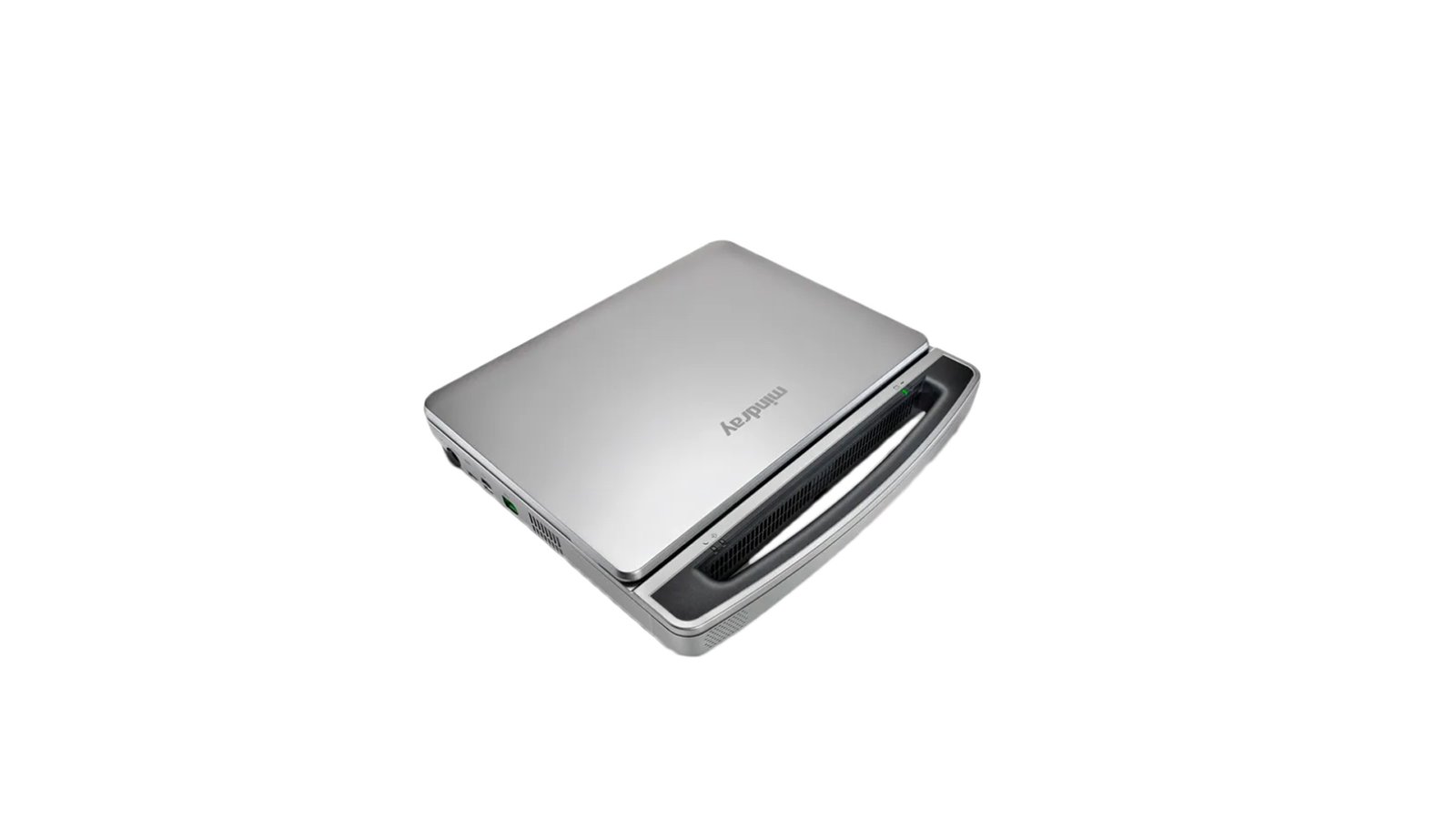
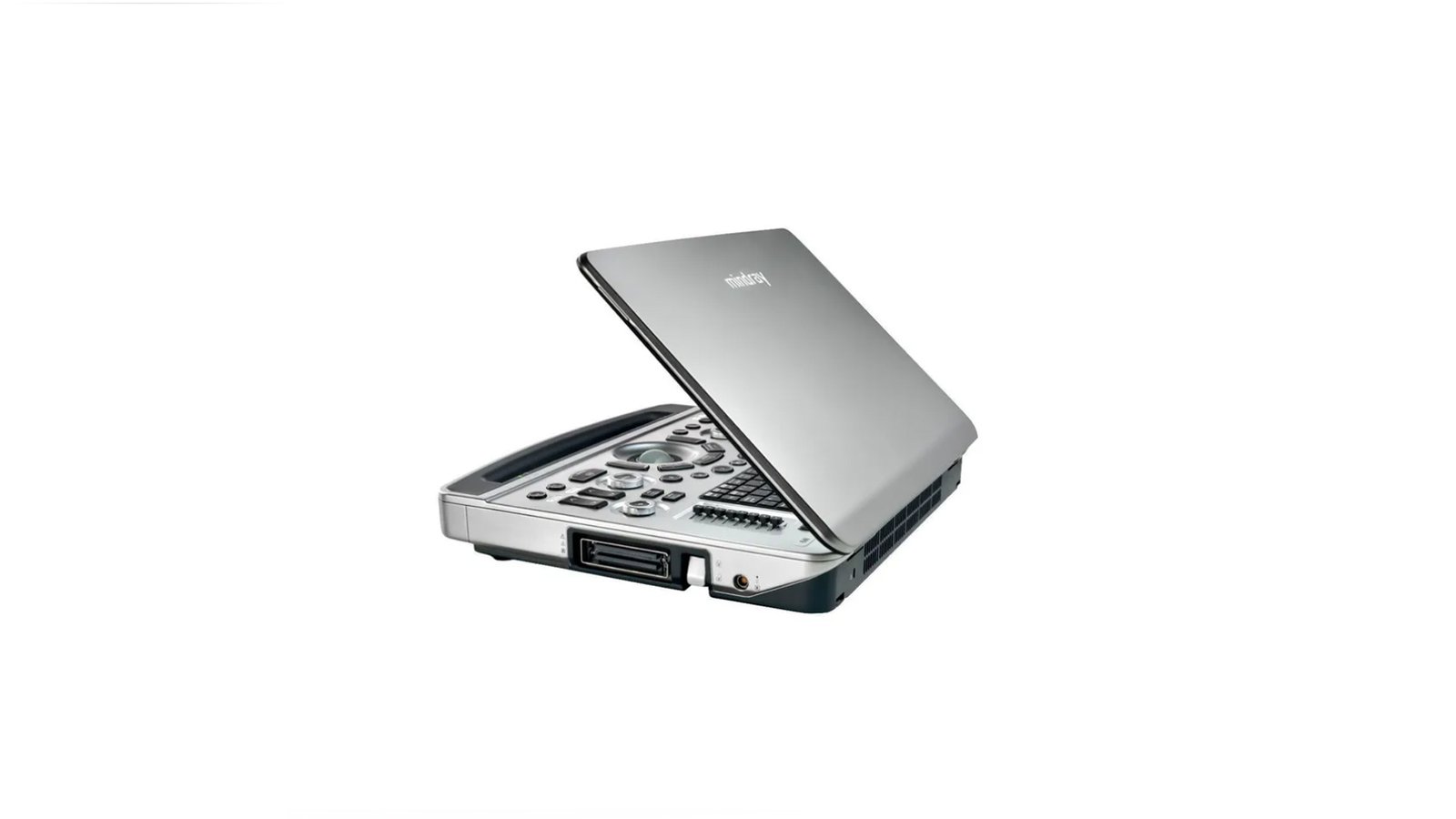
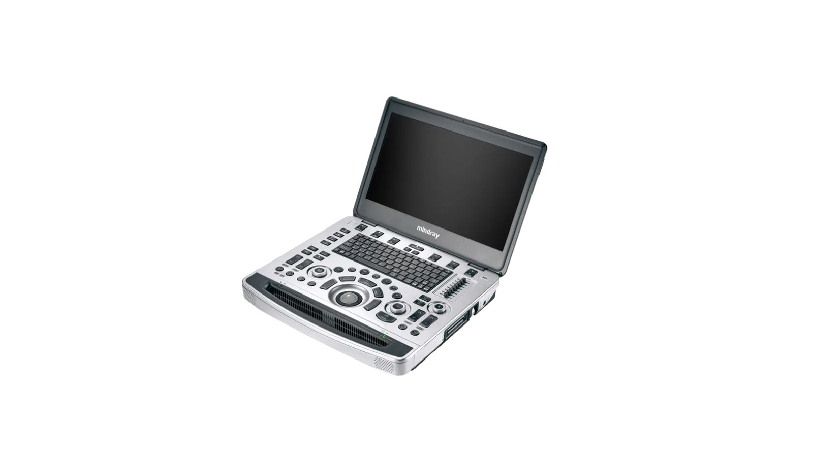
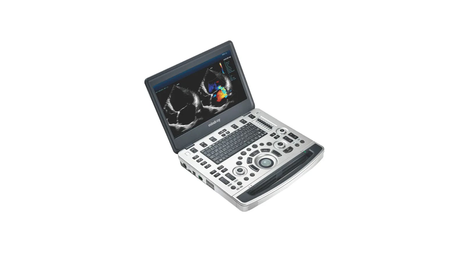
The Mindray M9T mobile ultrasound scanner can be used both as a portable ultrasound diagnostic device and together with a wheeled medical trolley. You can take this device to the patient's office, bring it to the hospital room or use it as a conventional stationary device. This ultrasound machine offers a wide range of capabilities, from elastrography to extended echo beamforming. All sensors compatible with the scanner are manufactured using Mindray's unique 3T technology to provide clearer imaging. Thanks to the additional use of single-crystal elements, the M9T has a higher penetrating power and improved color dynamics, which is especially important when scanning the so-called "hard-to-reach areas".
Functions:
Elastography Natural Touch
Natural touch elastography based on Mindray's latest patented technology makes examination results less dependent on the user's skills, thereby increasing examination accuracy in challenging clinical settings.
- Efficient seal detection
- Consistently high reproducibility of test results
- Imaging with UWN+ Contrast Agents (Nonlinear Ultra Wide Range Imaging)
The biggest advantage of the M9T ultrasound scanner is the support of Mindray's patented technology, which allows you to expand the possibilities of imaging with contrast. UWN+ technology enables the M9T to detect and exploit both secondary harmonics and non-linear primary signals, producing higher quality images. Higher sensitivity to minor signals, lower consumption of the active substance. Longer active agent action time and lower PSHI™ (Phase Shift Harmonic Imaging) measurement interval requirements. Isolated harmonic imaging for improved contrast resolution, delivering sharper images with superior spatial resolution and less noise.
Tissue harmonics - using additional harmonics generated in the tissues of the boundary layers, TG significantly increases the contrast resolution and improves image quality, especially when access to the study area is difficult.
TSV (Tissue Specific Imaging)
- Tissue-specific imaging optimizes image quality based on the properties of the tissue being examined. Four imaging options are available: general, muscle, liquid media, and adipose.
- iBeam - allows you to use multiple scanning angles to form a single image, resulting in increased contrast resolution and improved visualization.
- iClear - allows you to improve image quality based on automatic pattern recognition. Sharper edges and contours as well as smoother and more uniform tissue display. Grain reduction in "no-echo areas"
- Expanded Echo Beamformer - Expanded echo beamformer utilizes the traditionally ignored echoes of adjacent beams to form a single thinner, stronger image beam providing higher out-of-focus image resolution and deeper imaging penetration.
- Multi-beamforming - Up to 12x faster single-beam processing for superior temporal resolution and higher frame rates.
- Advanced Microflow Imaging - Allows better visualization of small vascular perfusions and better assessment of pathology perfusion by using a group of frames to generate merged images.
- iScape gives a complete and enhanced view of anatomical structures through panoramic imaging, combined with a speed indicator and a forward/reverse scan function, making the process easier, more consistent and more manageable.
- ExFOV - Get the most comprehensive diagnostic information at your fingertips with detailed anatomical imaging on all convex and linear transducers.
- B-Steer/ iNeedle is your tool for deeper biopsy: allows you to maneuver the ultrasound beam to improve the visibility of the needle, nerve fibers and small vessels.
- iStation, Mindray's unique patient information management system, allows you to seamlessly integrate, view, archive and retrieve patient data.
Execution of work operations:
- iZoom - provides instant switching to full screen mode with a single keystroke.
- Auto IMT - (automatic determination of the thickness of the intima-media complex) - automatic measurement of the thickness of the anterior and posterior walls, providing accurate information about the state of the carotid artery.
- iStation, Mindray's unique patient information management system, allows you to seamlessly integrate, view, archive and retrieve patient data.
- iTouch - allows you to perform instant automatic image optimization in B, color and pulsed wave Doppler (PW) modes with the press of a single key.
- iWorks is a high-tech tool that allows you to focus on the patient. Significantly reduces patient scan time through standardized procedures and user-configurable features.
- Raw Data Handling - Provides extensive post-processing options for saved images, including setting parameters, adding comments, and adding measurement data to ensure maximum productivity during scanning.
- Screen Size - ″15
- DICOM - Yes
- Command touch display - No
- The presence of compression elastography - Yes
- Availability of shear wave elastography - No
- Volumetric scanning (4D) - Yes
- 3D free hand reconstruction - Yes
- Automatic intima-media thickness (IMT) calculation - Yes
- Trapezoidal mode (virtual convex) - Yes
- Panoramic Scan - Yes
- The program for automatic measurement of the main parameters of fetal biometrics in obstetrics - Yes
- The presence of automatic calculation of the collar space - Yes
- Option Pack 5D - No
- Constant Wave Doppler (CW) - Yes
- Anatomical M-Mode - Yes
- Support for Fusion technology (combining images on CT / MRI with ultrasound) - No
- Volumetric imaging of the fetal heart (STIC) - No
- ECG Block Support - Yes
- Support for studies with contrast agents - Yes
- Device Type - Portable
- Specialization (ultrasound) - Cardiology
- Device class - Expert
- Type of elastography - Compression
- Types of supported sensors - Convex, Micro-convex, Pencil, Linear (up to 15 MHz), Linear low-frequency, Cavitary convex, Sector phased adult, Volumetric convex, Intraoperative linear, Sector phased pediatric, Sector phased neonatal, Transesophageal adult, Intraoperative convex, Linear ultrahigh frequency
- Supported Sensor Types - High Density, Monocrystalline, Matrix
- Number of active connectors for sensors - 1
- Gel warmer - No
- Color Doppler (CD) - Yes
- Tissue Doppler (TDI) - Yes
- Sensor splitter for portable scanners - Yes
- Built-in rechargeable battery - Yes
- Battery life of portable scanners (hour) - 1.5
- Assessment of myocardial deformation (speckle tracking) - Yes
- Solid State Drive (SSD) - Yes
- Height adjustable control panel - Yes
- Pulsed Wave Doppler Yes
- High Frequency Pulse Doppler - Yes
- Power Doppler - Yes
- Directed ED - Yes
- Vector Blood Flow Mapping - No
- Automatic determination of the Simpson ejection fraction - No
- Automatic Image Optimization - Yes
- Automatic calculation of hemodynamic parameters from the Doppler spectrum (tracing / spectrum contouring) - Yes
- Non-Doppler imaging of blood flow - No
- Stress echocardiography - Yes
- WiFi - Yes
- Needle Visualization Improvement for Linear Gauges - Yes
- Beam tilt in Doppler modes on linear B-steer transducers - Yes
- Multi-beam scanning/compounding - Yes
- Grain Suppression - Yes
- Virtual light source in 3D - No
- Option to obtain a three-dimensional image in the mode of color Doppler mapping (three-dimensional reconstruction of the color flow) - No
- Auto measurements in 3D mode - Yes
- High Sensitivity Doppler (Microvascular Imaging) - Yes