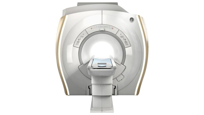
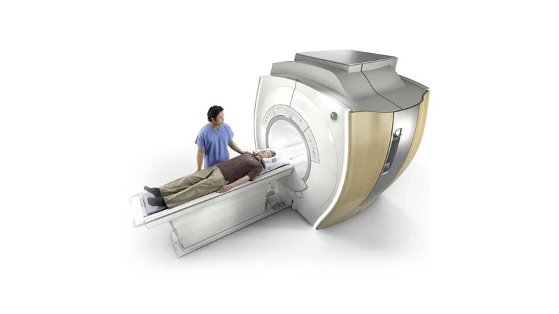
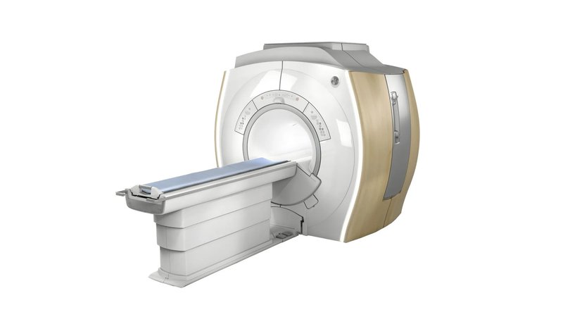
Магнитно-резонансный томограф GE Healthcare Optima MR360 Advance 1,5T
- Manufacturer
- GE
Optima MR360 Advanced
Built on the renowned high-definition platform used to develop GE's advanced MRI models. The equipment provides environmental safety for patients, versatility in use, high performance and image detail. One of the main advantages of the model is the quick readiness of the tomograph for almost any study of complexity. The modern system of the device provides high-quality visualization of various problem areas, such as blood vessels, chest and body. The Optima MR360 Advance is one of the safest and most energy efficient 1.5 Tesla (T) systems. Allowing you to use 34% less energy than previous generation scanners. And in combination with powerful software designed for 3 T tomographs, a balance is achieved between comfortable examination for patients and high quality of the images obtained. The Optima MR360 Advance has a large scan area (48 cm), which allows you to capture more anatomical zones with fewer scans. Thanks to the advanced development of General Electric, the equipment has a reliable magnetic field uniformity, 33/100 gradient and OpTix RF technologies. By equipping the workstation with software applications, doctors can process the data received from the tomograph in two-dimensional (2D) and three-dimensional (3D) planes.
Features
- Specialized programs for neuroimaging (three-dimensional visualization of the brain and spinal cord, examination of the state of blood vessels, etc.);
- Coil for examination of the brain and neck (examination of the vessels of the brain and neck, inner ear, temporal lobes, cervical spine, soft tissues, larynx and much more);
- Built-in coil for visualization of the cervical, thoracic and lumbar spine;
- Body coil for studies of the thoracic region, abdominal cavity, pelvic organs, vessels of the lower extremities and hip joints;
- Program for dynamic contrasting of the abdominal cavity and small pelvis (LAVA-Flex). Together with diffusion-weighted imaging, it is an essential tool in the diagnosis of oncology;
- Specialized coil for studies of the shoulder, elbow, wrist, knee and ankle joints, as well as the foot.
- FOV (field of view), cm - 50
- Aperture diameter, cm - 60
- Cardiac - Yes
- MR Angiography - Yes
- MR diffusion - Yes
- MR Perfusion - Yes
- MR Spectroscopy - Yes
- Maximum patient weight (kg) - 160
- Magnetic field strength, T - 1.5
- Motion Artifact Suppression Yes
- Acoustic Noise Reduction Yes
- Image Noise Reduction Yes
- Device type - High-field
- Contour type - Closed
- Magnet Type - Superconducting
- Cooling type - Liquid helium
- Functional MRI - Yes


