

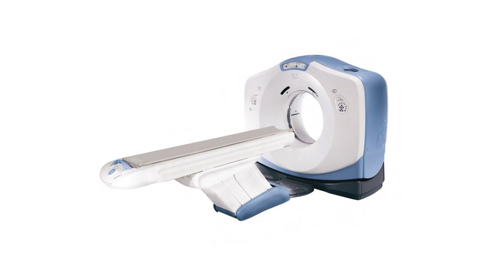


Компьютерный томограф GE Optima CT580
- Manufacturer
- GE
Computed tomograph GE Optima CT580
A modern innovative device for conducting an examination using x-rays to obtain a three-dimensional picture of the organ under study. For this purpose, X-ray imaging of the so-called sections of the organ with different exposures is carried out, after which computer processing of the obtained images forms a general three-dimensional picture of the organ under study. The GE Optima CT580 CT scanner is a 16-slice imaging system that provides exceptional image quality with significant spatial resolution. In this case, the value of the latter increases by 68% at the same radiation exposure. A slight degree of exposure is provided by the use of special technology. The GE Optima CT580 CT scanner is equipped with a detector with a wide field of coverage (50 cm), which provides the researcher with new clinical possibilities. The power of the X-ray generator (42 kW), the gantry aperture of 800 mm and the significant scanning range (170 cm) make it possible to examine even very obese patients (up to 227 kg). A modern X-ray tube does not require the equipment of a special cooling system, which makes it possible to carry out continuously even the most complex and lengthy manipulations. The GE Optima CT580 computed tomograph has a high spatial resolution matrix that visualizes the most complex organs from this point of view with the highest quality. The innovative technology provides a significant reduction in the degree of exposure and a digital signal. This reduction can be as high as 80% while maintaining the quality characteristics of the images. The GE Optima CT580 computed tomograph provides significant savings in contrast agent portions.
System Benefits
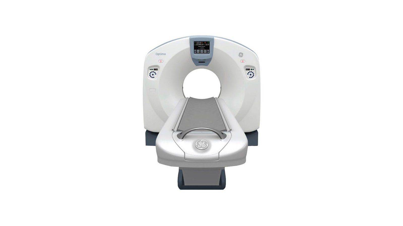
- GE Optima CT580 is a 16-slice CT that specializes in oncology diagnostics and planning the use of radiation therapy in the fight against tumors.
- Visualization of high-quality thin slices allows the system to pinpoint the exact location of the tumor for targeted application of radiation therapy.
- CT is equipped with a table designed for a large load capacity, which allows you to examine any patient without restrictions on body weight.
- The Optima CT580 features a wide aperture (80 cm) for maximum freedom in positioning large patients for complex exams.
- It is possible to perform CT colonography, a non-invasive procedure that is easily tolerated by patients, to detect polyps and colon cancer.
- New study protocols and expanded coverage make it possible to perform visceral perfusion studies to identify lung nodules and see how they develop over time, contributing to the effectiveness of treatment.
Virtual Simulation:
- determine the volume, contour and geometry of the beam automatically to increase the speed and accuracy of treatment plans during radiotherapy;
- 4D modeling can be carried out in 1 session, including an overview, contour and plan;
- less than 4 minutes will be required to contour about 15 structures with auto-segmentation of several organs.
- Metal Artifact Reduction technology significantly improves the quality of the resulting image, even if the patient under examination has metal objects (dental implants, joint prostheses), reducing the number of artifacts caused by metal.
Equipment:
- a table that is compatible with almost all devices for radiation therapy, and allows you to correctly position even patients with large weight;
- gantry with a wide aperture (65 cm) and an increased field of view makes it possible to easily position oversized patients in complex studies;
- MicroVoxel is a feature for isotropic fine detail imaging that provides accurate contouring for an accurate treatment plan;
- Advantage 4D is a feature that synchronizes CT with the patient's respiratory movements, which reduces structural deformations and determines the dynamics of movement for an accurate assessment of treatment planning.
Gantry
- Aperture: 80 cm;
- Remote tilt gantry function from the operator's console;
- Built-in "breath instructor" and countdown timer;
- Built-in scan start button with X-ray countdown timer;
X-ray tube
- Heat capacity 8 MHU;
- Small focal spot: Nominal focal spot size 0.7 x 0.6 mm (IEC60336/1993);
- Small focal spot: Nominal focal spot size 0.7 x 0.6 mm (IEC60336/1993);
- Large Focal Spot: Nominal focal spot size 0.9 x 0.9 mm (IEC60336/1993).
Standard Functions and Features
- IQ Enhance (IQE) is a special algorithm designed to reduce the number of artifacts in helical scan mode in thin slices. This algorithm allows, by increasing the scanning pitch, to cover more anatomical areas of the body with the same image quality;
- Chest Kernel makes it possible to reconstruct the details of the lungs and the structure of the mediastinum;
- Dose Check compares the planned exposure dose with the allowable exposure dose;
- AutomA/SmartmA: Automatic current optimization function (mA) to maintain a constant noise level in the image under different collimation/detector configurations, scan modes, scan rotation speed, table speed, slice thickness. When changing parameters, the scanning technique is adjusted;
- SMART PREP: software for real-time monitoring of contrast enhancement and automatic playback of previously recorded messages after switching the scanning phase by the operator. With the Auto Minimum Delay function, the positioning time is automatically set to the minimum;
- Helical Tilt is located on the operator's console and serves to increase the productivity of the study, includes built-in accessories that help avoid injury when the patient interacts with the CT scanner gantry;
- Large Image Series allows you to store up to 3000 images in one sequence;
- Data Export is a standalone tool for converting clinical images into computer compatible formats (jpeg, mpeg, avi). This tool allows you to create more convenient reports for both doctors and patients;
- CD/DVD Interchange: Write images to CD-R or DVD-R via DICOM Interchange using DICOM 3.0 format;
- Direct 3D creates a 3D model during axial reconstruction of the image, reducing the need for user intervention in the process;
- Wideview: Display Field of-View (65 cm) is a proprietary algorithm for generating an approximation of the CT image reconstruction outside the field of view;
- DMPR: DMPR function with Auto-Batch mode, allows automatic real-time reconstruction and transmission of multi-planar images, in any plane.