

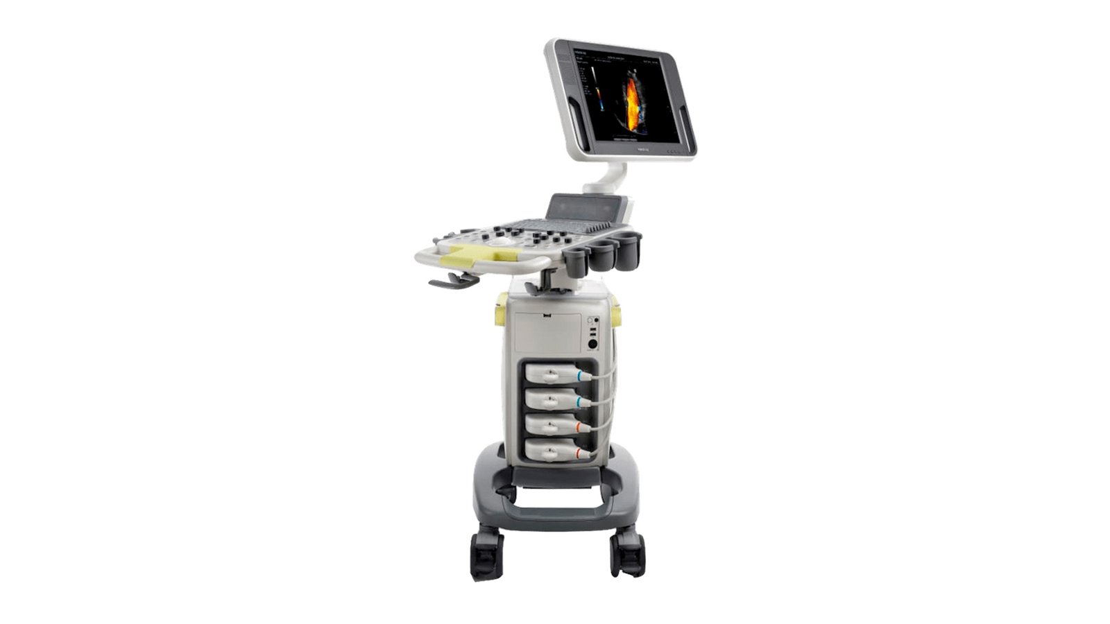
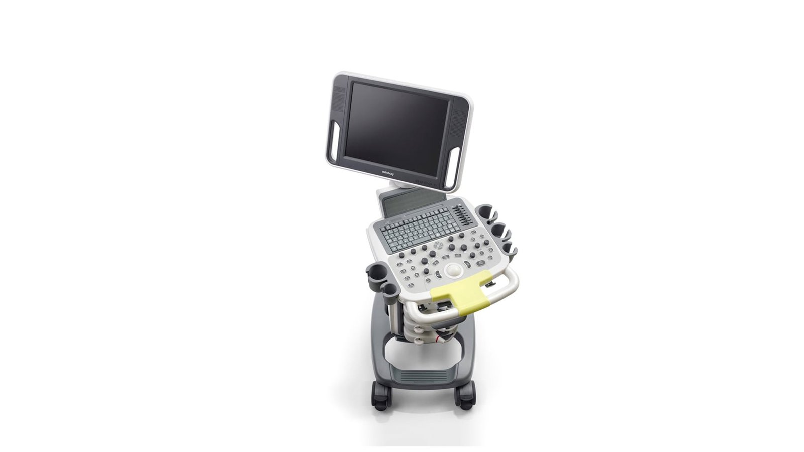
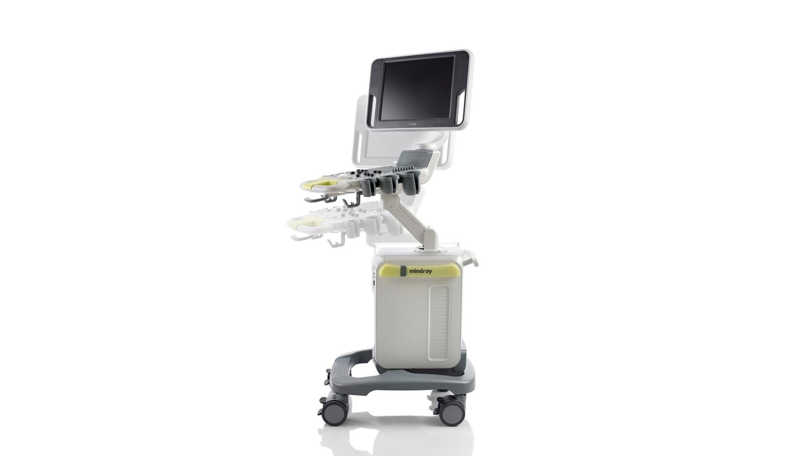
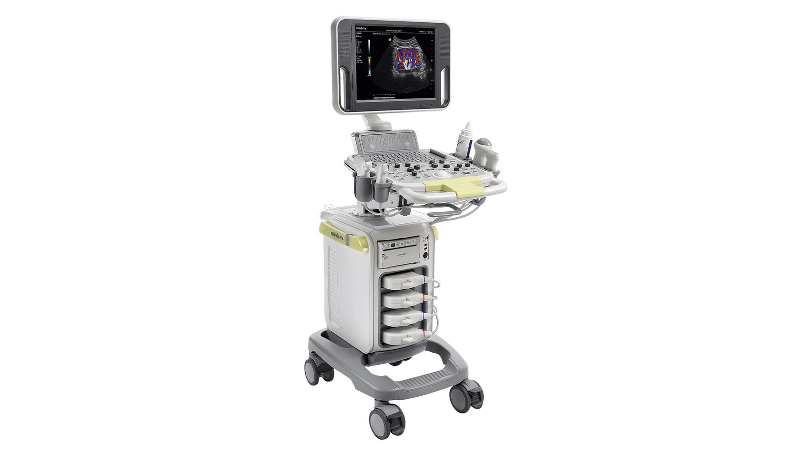


Аппарат УЗИ Mindray DC-N3
Ultrasound scanner DC-N3
Representing the ultimate combination of quality, versatility and affordability, it truly redefines fundamentals, delivering best-in-class functionality and performance at a reasonable price.
PSH (Phase Shift Harmonic Imaging) Functions
Isolated harmonic imaging for improved contrast resolution, delivering sharper images with superior spatial resolution and less noise.
iBeam
Allows multiple scanning angles to form a single image resulting in increased contrast resolution and improved visualization.
iClear
Allows you to improve image quality based on automatic pattern recognition.
Sharper edges and contours
Smooth and uniform tissue display
Reduced graininess in "areas without echo"
Multi-beam shaping
Up to 4x faster single-beam signal processing for superior temporal resolution and higher frame rates.
iScape
Gives a complete and enhanced view of anatomical structures through panoramic imaging, combined with a speed indicator and a forward/reverse scan function, making the process easier, more consistent and more manageable.
ExFOV
You will have the most complete diagnostic information at your disposal thanks to detailed visualization of anatomical structures on all convex and linear transducers.
b-steer
Your tool for deeper biopsy: maneuvers the ultrasound beam to improve visibility of the needle, nerve fibers and small vessels.
Free Xros M
Allows you to get accurate anatomical measurements by freely placing M-mode lines at any angle. Optimum image quality is achieved by using up to 3 M-mode lines at the same time.
Free Xros CM
Allows you to get complete information about the movement of the heart muscle in different phases of contraction and at the same time determine the degree of synchronization of the myocardium. The accuracy of the results is ensured by the high frame rate.
Tissue Doppler Imaging (TDI)
A study in tissue Doppler mode allows you to determine quantitative indicators of the movement and functioning of the heart muscle, provides a full range of tissue Doppler modes to reduce time and increase the accuracy of ultrasound diagnostics.
Execution of work operations
iStorage / iMeasurement / iReport
iStorage: direct transfer of images and reports to a PC via a network cable.
iMeasurement & iReport: offline PC software for user defined measurement table, calculation formulas and report templates.
Auto IMT (automatic determination of the thickness of the intima-media complex)
Automatic measurement of the thickness of the anterior and posterior wall, providing accurate information about the state of the carotid artery.
Auto LV
Simple left ventricular measurement with automatic ejection fraction tracking and manual adjustment.
iTouch
Enables instant automatic image optimization in B, Color and PW modes at the touch of a button.
iZoom
Provides instant switching to full screen mode with a single keystroke.
iStation
Mindray's unique patient information management system allows you to seamlessly integrate, view, archive and retrieve patient data.
Working with "raw data"
Opens up a wide range of post-processing options for saved images, including setting parameters, adding comments and measurement data, which ensures maximum productivity during scanning.
- DICOM - Yes
- WiFi - Yes
- Auto measurements in 3D mode - No
- Automatic Image Optimization - Yes
- Automatic calculation of hemodynamic parameters from the Doppler spectrum (tracing / spectrum contouring) - Yes
- Automatic intima-media thickness (IMT) calculation - Yes
- Automatic determination of the Simpson ejection fraction - No
- Anatomical M-Mode - Yes
- Vector Blood Flow Mapping - No
- Type of elastography - Compression
- Types of supported sensors - Convex, Micro-convex, Pencil, Linear (up to 15 MHz), Linear low-frequency, Cavity convex, Cavity biplane (convex + convex), Sector phased adult, Volumetric convex, Linear high-frequency
- Virtual light source in 3D - No
- Built-in rechargeable battery - Yes
- High Frequency Pulse Doppler - Yes
- High Sensitivity Doppler (Microvascular Imaging) - No
- Pulsed Wave Doppler - Yes
- Device class - Medium
- Number of active connectors for sensors - 3
- Command touch display - No
- Multi-beam scanning/compounding - Yes
- Beam tilt in Doppler modes on linear B-steer transducers - Yes
- The presence of automatic calculation of the collar space - Yes
- The presence of compression elastography - Yes
- Availability of shear wave elastography - No
- Directed ED - Yes
- Non-Doppler imaging of blood flow - No
- Volumetric imaging of the fetal heart (STIC) - No
- Volumetric scanning (4D) - Yes
- Option to obtain a three-dimensional image in the mode of color Doppler mapping (three-dimensional reconstruction of the color flow) - No
- Assessment of myocardial deformation (speckle tracking) - No
- Option Pack 5D - No
- Panoramic Scan - No
- Grain Suppression - Yes
- ECG Block Support - Yes
- Support for studies with contrast agents - No
- Support for Fusion technology (combining images on CT / MRI with ultrasound) - No
- Gel warmer - No
- Constant Wave Doppler (CW) - Yes
- The program for automatic measurement of the main parameters of fetal biometrics in obstetrics - Yes
- Sensor splitter for portable scanners - No
- Screen Size - ″17
- Height adjustable control panel - Yes
- Specialization (ultrasound) - General studies
- Stress echocardiography - No
- Solid State Drive (SSD) - No
- Device type - Stationary
- Supported Sensor Types - High Density
- Tissue Doppler (TDI) - Yes
- Trapezoidal mode (virtual convex) - Yes
- 3D reconstruction by "free hand" method - No
- Needle Visualization Improvement for Linear Gauges - No
- Color Doppler (CD) - Yes
- Power Doppler - Yes