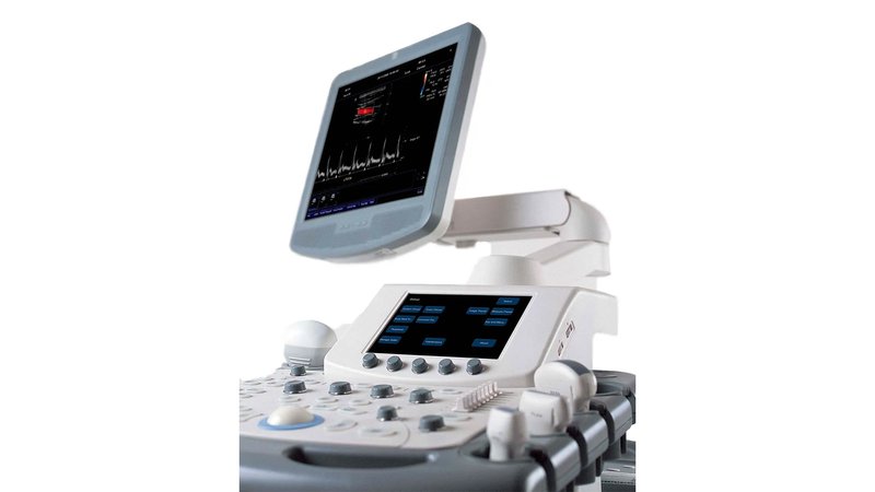
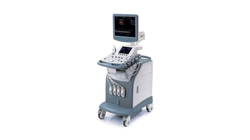
Узи аппарат Mindray DC-7
- Manufacturer
- Mindray
Description
Mindtay DC-7
Diagnostic ultrasound scanner for a wide range of studies from Mindray. It belongs to high-class devices and is used in hospitals, medical centers and antenatal clinics. Widespread use of the Mindray DC 7 ultrasound scanner makes it possible to conduct examinations of all organs and systems of the body (from examination of the vessels of the head and neck, abdominal examinations, to gynecological and urological examinations), makes it possible to work with difficult patients, also visualizing well.
Scanning
- Possibility of 4D images with volumetric visualization of the fetus or organs in real time for accuracy and speed in making a diagnosis or determining the duration of pregnancy;
- Simultaneously 2 or 4 sonograms in B-mode;
- High photo quality in M-mode due to the formation of a thin and strong beam (M mode is combined with B);
- Trapezoidal imaging is performed on linear sensors, which will expand the field of view;
- TDI (tissue doppler) for visualization of the movement of tissues with doppler and includes different types of scanning, including color;
- Panoramic images appear with iScape;
- 3D reconstruction is possible in Smart3D;
- A program with which you can conduct research with contrast agents;
- B&W and color images can be displayed on the screen.
Functions
- Keeping film "on hand" is no longer relevant, with the new DC-7 feature, images are transferred to a USB drive for quick file review and without the possibility of "paper obsolete" when the pictures were printed. It also reduces the cost of consumables for the product;
- Since not everyone can use a USB file system, in this case, printing of images on a printer, recording to a disk or cassette is provided;
- Ultrasonic scanner Mindray DC-7 supports the DICOM 3.0 program - this is the creation, saving, transfer and display of patient data;
- Different scaling of the image will not let you miss any change in organs and systems, and the convenience of a large screen helps to perform the study more quickly;
- Power save mode extends equipment life and extends equipment maintenance intervals;
- CW (constant wave doppler) - assessment of high velocity blood flow in the vessels;
- The iBeam function sharpens and defines the edges of the image, reduces the artifacts found by the device, and increases the contrast;
- Has a backlit keyboard on the panel;
- iClear (speckle noise reduction) technology reduces graininess in images for improved evaluation and diagnosis;
- iVision has a slideshow function, and a convenient catalog-style menu customizes the search and makes it easier and faster;
- Q-Click - the ability to quickly adjust the parameters that are displayed on the screen;
- The range of sensors increases the clinical application of research equipment;
- Linear transducers increase the field of view during an ultrasound examination and provide more information for clinical evaluation;
- The presence of THI (tissue harmonization) increases penetration into tissues, which will help not to miss the pathological process;
- Duplex mode - visual assessment of vascular patency, identifying the cause of disorders (stenosis, atherosclerotic plaques, developmental pathologies), as well as studying the speed and direction of blood flow;
- Triplex mode - will expand the capabilities of duplex by adding CD mode. The ability to study the direction of blood flow and its speed for the presence of stenosis or its degree;
- Stress-Echo-KG - signs of cardiac ischemia provoked by physical activity or farm are examined. drugs.
Supported sensors
- Convex;
- Microconvex;
- Sector phased;
- Linear;
- Combined rectovaginal;
- Biplane endorectal;
- Specialized;
- 4D sensors;
- Multi-frequency, high-density broadband.