УЗИ аппарат Mindray DC-55
Description
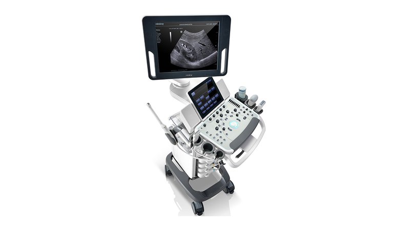
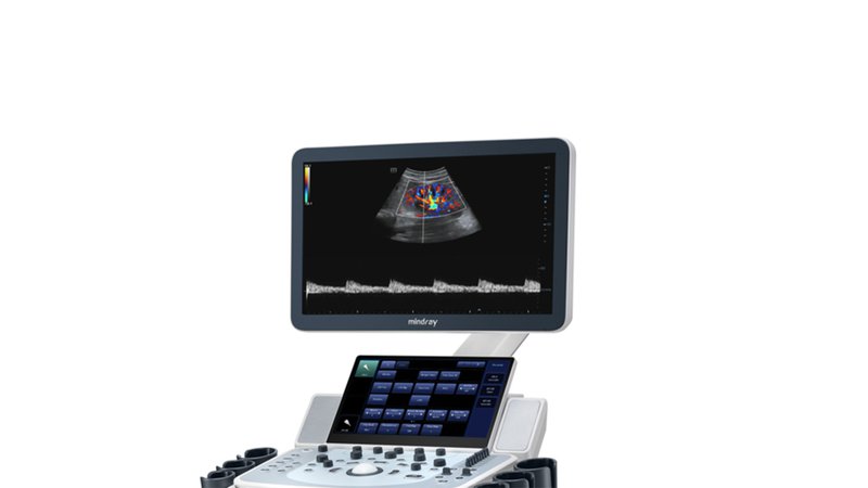
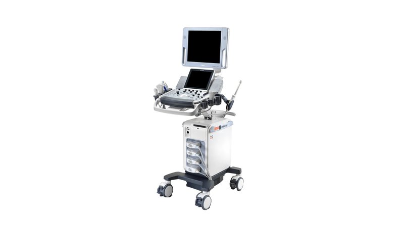
Ultrasound machine Mindray DC-55
Committed to providing quality and affordable medical care, Mindray has developed the DC-55 next generation multifunctional ultrasound system. The DC-55 Ultrasound Scanner combines an application suite, automated measurement tools, and built-in tutorials to make ultrasound examinations accurate, efficient, and affordable, and provide users with an exceptional experience. Mindray understands the importance of optimal performance. The 10.4-inch touchscreen provides ease of use and helps ease the burden of daily clinical work, while the compact design and built-in battery ensure maximum portability.
Basic configuration:
- 17" high resolution LED monitor
- Command touch screen 10.4" with gesture recognition technology and the ability to adjust the angle
- Scan Modes B/M/Color Doppler CDI/Color M/Power Doppler PD/Dir.PD Power Doppler
- PW Doppler (including high pulse repetition rate mode HPRF)
- PSH™ - phase inverted harmonic
- iBeam™ - multibeam compounding mode
- iClear™ - adaptive noise reduction mode
- iTouch™ - automatic image optimization
- iZoom™ - Full Screen Ultrasound View
- Echo Boost™ - enhanced imaging mode for cardiology
- HR Flow - blood flow display mode with high temporal and spatial resolution for accurate and uniform visualization of vessels, including the smallest
- Saving information in raw data format
- Shared Service Package - preset parameters, annotations, markers, measurement programs for abdominal, obstetrics, gynecology, cardiology, angiology, small parts, urology, pediatrics, emergency medicine
- 500 GB hard drive with iStation™ patient database software
- DVD-RW drive
- 4 connectors for connecting sensors (standard connection scheme: 3 regular + 1 high-density)
- HDMI output and USB 3.0 ports
- MedSight™ - transfer of information to the patient's electronic devices (available for IOS / Android operating systems, the DICOM Basic option on the ultrasound scanner is required to work with IOS devices)
- MedTouch™ - scanner control from doctor's electronic devices (available for IOS/Android devices)
- iScanHelper - built-in training software
- Holder for intracavitary probe (by default on the left side of the scanner, if you need its location on the right side - select the "Right" option before ordering)
Possible hardware and software options:
- Physio Module - IEC standard physiological signal recording module
- CW - block of constant wave doppler
- 4D - real-time volumetric scanning unit
- Battery - built-in rechargeable battery
- Built-in Wireless Adapter - built-in adapter for wireless data transfer
- Smart OB™ is an automatic calculation program with the ability to manually edit the main obstetric indicators: BPR, BP, OH, LZR, using algorithms for automatic contouring and recognition of organ boundaries
- Smart NT is a program for automatic determination and calculation of the thickness of the nuchal space in the fetus
- Smart 3D™ – freehand 3D ultrasound imaging without the use of bulk transducers
- iLive™ - 3D imaging mode using virtual light-shadow processing technology with the ability to move the light source (requires Smart 3D option or 4D module)
- iPage™ - multislice tomographic display with adjustable slice thickness (requires 4D module)
- iNeedle™ - improved visualization of needles during biopsy with linear probes
- iScape™ View - panoramic scanning - displaying objects of great extent on the screen (simultaneous display of large volumetric formations, structures and organs on the screen over a large area, etc.) with the possibility of performing calculations and measurements
- iWorks™ - automated workflows for all major exam types
- Auto IMT Package - measurements and analysis of the thickness of the intimamedia complex (IMT) of the carotid artery
- Natural Touch Elastography - an option for assessing tissue elasticity (elastography), with an analysis program. Valid on linear probes 7L4A and L14-6NE
- Free Xros M ™ - anatomical M-mode is the ability to rotate the cursor in M-mode at an arbitrary angle (with a fixed sensor position) and, accordingly, obtain a graph of the movement of heart structures in various arbitrary planes
- Free Xros CM™ Anatomical Envelope M-Mode (Requires Option TDI)
- TDI (Tissue Doppler imaging, including TDI Color, Power, PW and M mode)
- TDI Quantification Analysis Software - Quantitative Tissue Doppler Analysis (Requires Option TDI)
- Stress Echo - a package for conducting and evaluating