УЗ система Mindray DC-70 Pro X-Insight
Description
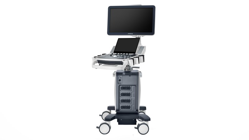
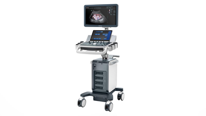
Ultrasound machine Mindray DC-70 Pro X-Insight
The Mindray DC-70 Pro X-insight is an expert-class ultrasound scanner equipped with a GPU and CPU, which provides multi-core parallel processing for fast image acquisition. With an advanced engine, the image processing speed is three to four times faster than other technologies, providing incredibly fast image processing for 3D/4D and other applications.
Features
- X Engine
The X-Engine is equipped with a GPU and a CPU to provide multi-core parallel processing for fast image acquisition. With an advanced engine, the image processing speed is three to four times faster than other technologies, providing incredibly fast image processing for 3D/4D and other applications.
- New version of iLive
Comprehensive enhancements to iLive dramatically improve image resolution and anatomical fidelity. Hyaline is a new imaging technique that dynamically applies transparency to displayed structures to better reproduce anatomical features and better show interiors close to hard surfaces.
- Monocrystalline sensors with 3T technology
Combined with Mindray's 3T technology (triple matching layers, fully separated crystals, thermal control), the new single crystal elements, linear and phased scan sensors provide wider bandwidth, deeper scanning and higher resolution. The optimal solution for scanning in gynecology, abdominal surgery, cardiology, etc.
- Combo Wave Sensors
Compared to traditional transducers, ComboWave incorporates a new type of complex piezoelectric material that dramatically improves the acoustic spectrum and reduces acoustic impedance. Incorporating Mindray's unique 3T technology, ComboWave linear probes provide excellent performance and exceptional image resolution and uniformity for scanning thyroid, chest, blood vessels, and more.
- Smart FLC
Automatic quantification and volume calculation of follicles from a 3D volumetric image.
- Smart Planes CNS
The tool greatly improves scanning accuracy, combined with fully automated operation, resulting in accurate diagnostics, improved throughput, and reduced user dependency.
smart face
- Smart Face
provides fast, intelligent image optimization of the fetal face with a simple push of a button. The system can immediately remove obstructions such as the umbilical cord, placenta, uterus, and extremities in volumetric data and create an optimal image of the fetal face with minimal effort.
- Smart OB
Automatic fetal measurement software: measure and calculate Bipariental Head Size (BPD), Fronto-occipital Head Size (OFD), Head Circumference (HC), Abdominal Circumference (AC) and Femur Length (FL) at the touch of a button.
- Smart NT
Automatic measurement of the thickness of the collar space.
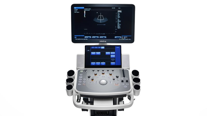
- Full HD monitor
Mindray's unique monitor technology provides an unlimited angle of motion for a floating design for comfortable monitor positioning according to clinical needs. Diagonal 21.5/23.8 inches.
- Ultra-thin touch screen
Diagonal 13.3 inches and tilt adjustment. Gesture control sets new trends in the design of portable ultrasound systems, providing a fast, intelligent and intuitive workflow that exceeds any expectations.
Available options :
- Physiological signal recording module (includes ECG and FCG) of the AHA standard;
- Physiological signal recording module (includes ECG and FCG) IEC standard;
- 4D and TEE Module for connecting volumetric and transesophageal sensors;
- Block of constant-wave Doppler;
- Built-in battery (provides 24 hours of keeping the scanner in sleep mode);
- Mode for automatic contouring, counting and sizing of follicles from a volumetric image (requires a 4D/TEE model);
- The program for automatic measurement of the main parameters of fetal biometrics in obstetrics (Requires a package of calculations for obstetrics Obstetrics);
- Program for automatic determination and calculation of the thickness of the nuchal space in the fetus (Requires the software package for obstetrics Obstetrics);
- Three-dimensional reconstruction by the "free hand" method;
- 3D rendering mode using virtual light-shadow return technology with the ability to move the light source (requires 4D/TEE module);
- iPage function - multislice tomographic display with adjustable slice thickness (requires 4D/TEE module);
- Obtaining an arbitrary slice of a given thickness in a three-dimensional image with simultaneous improvement of contrast, including along a derivative curve (4D/TEE module is required);
- Software module for obtaining a volumetric image of the fetal heart with high temporal and spatial resolution in gray scale and color flow modes (4D / TEE module is required);
- Color Doppler / Power Doppler 3D imaging option (requires 4D/TEE module);
- Arbitrary selection of a scanning slice in the obtained volumetric image with simultaneous display of three planes (requires the 4D/TEE module);
- Program for automatic calculation of the volume and dimensions of structures obtained by volumetric echography (requires the 4D/TEE module);
- Tissue elasticity assessment option (elastography), with analysis program (supported on L12-3E, L9-3E, L14-6WE, L14-6NE, V11-3E, V11-3BE, V11-3HE, DE11-3E sensors);
- Contrast agent option (supported on SC6-1E, C5-1E, C5-2E, L12-3E, L14-5WE, L9-3E, V11-3E, V11-3HE, DE11-3E transducers);
- Package for quantitative analysis when conducting surveys using contrast agents (requires the UWN Contrast Imaging option);
- Needle visualization enhancement option for linear gauges;
- Software module for obtaining panoramic images;
- Automated work protocols for all major types of research;
- Automatic calculation of the thickness of the intima-media complex with the analysis program (requires the package of vascular calculations Vascular);
- Anatomical M-mode (up to 3 slices);
- Tissue Doppler including color mapping, pulsed tissue doppler, power tissue doppler and tissue M-mode;
- Enveloping anatomical M-mode (requires installed TDI option);
- Tissue Doppler quantification software (requires TDI option installed);
- A package for conducting and evaluating the results of stress echocardiography (requires a module for recording physiological signals of ECG / PCG Physio Module);
- Package for quantitative assessment of movement and deformation of the myocardium based on registration of the displacement of segments of the myocardium of the heart (requires the input module for physiological signals ECG / PCG Physio Module);
- Left ventricular imaging software package with contrast (available on SP5-1E, P4-2E transducers).
Connecting to a DICOM network
- Basic - a basic set of options: saving to the server and media, printing
- Worklist - option to download a list of tasks from the server
- MPPS - an option that allows you to upload additional information about the conditions of the survey to the server
- Query/Retrieve - request and retrieve patient data and images from the server
- Cardiac S/R - structured cardiology report
- Vascular S/R - Structured Angiology Report
- Ob/Gyn S/R - OB/GYN Structured Report
- Breast S/R - structured breast examination report
Areas of use:
- Abdominal examinations
- obstetrics and gynecology
- Cardiology
- Vascular Research
- Neurology
- Traumatology and Orthopedics
- Urology
- Endocrinology
- Pediatrics
- Neonatology
- Transcranial studies
- Oncology