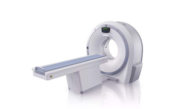
Компьютерный томограф GE Healthcare Revolution АСТ
- Manufacturer
- GE
Description
Computed tomograph GE Healthcare Revolution ACT
Revolution ACT - Produces high quality images and reliably visualizes anatomy in 3D with the Clarity detector providing 18 lp/cm spatial resolution. Visualization of all structures of the heart in one cardiac cycle with high resolution and suppression of artefacts of the movement of the coronary arteries at any heart rate. Scans to assess coronary atherosclerotic changes or a comprehensive cardiac assessment can be performed without the use of beta-blockers. The Gemstone Clarity detector provides scanning with a coverage area of 160 mm. Possibility to open many sessions for one or more patients at the same time.
Features
- Reducing the radiation dose by changing the anode current of the X-ray tube for surface tissues;
- Before scanning, the doctor can choose the required ratio of noise level and image quality: after that, the real-time CT scanner will automatically select the exposure parameters for a particular patient and a particular scan;
- 3D filters for noise reduction;
- The device allows you to control the x-ray beam in real time;
- Algorithm for reducing artifacts;
- Sagittal, coronal, oblique and curvilinear modes are available;
- Smart Dose Technologies:
- ViSR Volumetric spatial reconstruction of the image;
- ODM The technology is designed to perform the function of a virtual shield;
- DOSE CHECK Dose warnings;
- 3D mA MODULATION Optimizes tube current in the x-y-z directions with little impact on image quality;
- DOSEWATCH™ The program ensures the maintenance of the minimum acceptable level (ALARA) of radiation exposure.
Workflow management
Starting and ending an exam is faster than normal CT exam mode. Information about the patient is pulled up automatically.
Starting a study to achieve a given density of contrast agent.
During the study, images are displayed in real time. As soon as the edge of the desired anatomical zone is reached, the study can be stopped. How to reduce unnecessary radiation dose for the patient.
Possibility to open many sessions for one or more patients at the same time. In parallel, film installation is available.
Radiation exposure management
Reducing radiation dose by modulating the anode current of the X-ray tube for surface tissues such as mammary glands, lens, etc. Pixel noise averaging without reducing efficiency. Reduction of radiation exposure up to 40% is achieved
Custom tools for dose management in clinical practice and based on the XR-25-2010 standard published by the Electrical and Medical Equipment Manufacturers Association
Before scanning, the doctor can select the required ratio of noise level and image quality: after that, the real-time CT scanner will automatically select the exposure parameters for a particular patient and a particular scan
3D filters to reduce noise (standard deviation) and enhance brain, tumor and pediatric imaging. 20% improvement in image quality for the same patient dose or 36% reduction in dose for the same image quality
Real-time X-ray beam tracking technology that guarantees high resolution without dose escalation and collimation on the patient outside the exam
Protocols - pediatric protocols, for conducting pediatric research and reducing medical errors
The short gantry geometry in combination with the generator and tube allows efficient use of X-rays
Special algorithm to reduce artifacts that appear during spiral research
working platform
Multiplanar reconstruction provides the ability to study anatomy in real time in curvilinear modes. Sagittal, coronal, oblique and curvilinear modes available
Multiplanar volumetric reconstruction is a quick and easy way to generate volumetric images for CT angiography without setting data thresholds or eliminating unwanted anatomy
Direct multiplanar reconstruction mode with support for automatic batch processing, providing automatic real-time direct reconstruction and transmission of fully corrected multiplanar images in any plane;
Prospective Multiple Reconstruction - Up to 10 sets of reconstructions can be programmed as part of the study protocol. The operator can select different start and end points, slice thickness, spacing, reconstruction algorithm and display field size for each reconstruction;
post-processing
Volume analysis allows you to manage 3D and 2D images in real time. Multiple series can be viewed on one display by merging 2D images. Virtual endoscopy allows visualization of