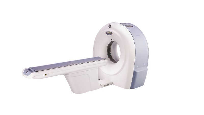
Computed tomograph GE Healthcare Brivo CT325
- Manufacturer
- GE
Description
Computed tomograph GE Healthcare Brivo CT325
A modern innovative device for conducting an examination using x-rays to obtain a three-dimensional picture of the organ under study. For this purpose, X-ray imaging of the so-called sections of the organ with different exposures is carried out, after which computer processing of the obtained images forms a general three-dimensional picture of the organ under study. The GE Brivo CT325 CT scanner is a 2-slice imaging system that provides exceptional image quality with significant spatial resolution. In this case, the value of the latter increases by 68% at the same radiation exposure. The GE Brivo CT325 CT scanner is equipped with a 20 mm detector, which provides the researcher with new clinical possibilities. The power of the X-ray generator (24 kW), the gantry aperture of 70 cm and the significant scanning range (120 cm) make it possible to examine even very obese patients (up to 180 kg). A modern X-ray tube does not require the equipment of a special cooling system, which makes it possible to carry out continuously even the most complex and lengthy manipulations. The GE Brivo CT325 computed tomograph has a high spatial resolution matrix that visualizes the most complex organs from this point of view with the highest quality. The GE Brivo CT325 computed tomograph provides significant savings in contrast agent portions.
Features and advantages
Volume Analysis function
Allows you to work with 3D and 2D images in real time. On one display, you can see many raw images, 2D images associated with different slice thicknesses (Average, MIP, MinIP, Volume Rendering) as required by the study.
Reformatting
-Multi-Planar Reconstruction (MPR) presents datasets as they would appear in the axial, sagittal, coronal, and oblique planes to diagnose sinus, thoracic, abdominal, internal disc, and fracture pathologies.
Multiplanar Volumetric Reconstruction (MPVR)
Allows you to quickly and easily create volumetric images to help you gently increase contrast and improve visualization of structures. MIP (Maximum Intensity Projection) functions for vascular anatomy; MinIP (Minimum Intensity Projection) can be applied to the airways and bronchi; function Average (Average) - for studies of the head and abdominal organs. The selected volume can be viewed from any selected plane in combination with varying slice thickness to provide a clearer view of the pathology of the pancreas, renal arteries and spine.
3D surface, 3D MIP and 3D volumetric reconstruction
3D rendering enhances the 3D rendering or rendered image with transparent rendering. This reconstruction provides more information about the spatial relationships of various structures in comparison with the standard surface 3D reconstruction. So you can accurately and confidently interpret CT scans.
Virtual Endoscopy
Provides visualization of intraluminal structures such as airways, sinuses, or vascular structures. Images can be viewed dynamically using the "fly through" mode.
CT Perfusion
Ability to manage patients with cerebral stroke through rapid quantitative analysis of blood disorders in the brain, including cerebral blood volume, cerebral blood flow and mean plasma residence time.
CT colonography
The ability to quickly, accurately, non-invasive examination of the colon. The prone and supine images can be displayed and synchronized together. It is also possible to even "fly" between 3D structures, which will be reminiscent of an optical colonoscopy. The ability to leave marks, for example, where a polyp was found, in addition, there are tools to assess the distance and area of interest for size and homogeneity.
Vascular analysis
Allows for in-depth analysis of vessel properties with automatic determination of the center line of the vessel. Tracking multiple paths to measure angles between vessel branches. It is also possible to view true oblique sections of vessels, rotate non-linear views for a clearer visualization of vascular lesions.
DentaScan function
Possibility to create full-fledged sets of complex cross reformations of the upper and/or lower jaw in the axial, oblique plane and in the panorex projection. The DentaScan feature provides information needed for prosthetic planning or orthodontic surgery.